Solitary cutaneous mastocytoma
Solitary cutaneous mastocytoma
This is a solitary lesion that occurs almost exclusively in children with no obvious site predilection, although a study suggested an affinity for the extremities (Fig. 29.347). It accounts for 10–20% of cases of mastocytosis.24,38–41 Lesions are nodular with a reddish or yellow color, and usually do not exceed 1 cm in diameter.4,24,38–40 They may be associated with flushing attacks.13,39 Most cases resolve spontaneously, whilst the few cases that persist are cured by excision.4,11,35,38
Constitutional symptoms of fatigue, weight loss, fever, and musculoskeletal complaints including bone pain, osteopenia, osteoporosis, fractures, arthralgias, and myalgias may be seen. Minimal splenomegaly is sometimes seen, and rarely there may be lymphadenopathy and hepatomegaly.4,8,18,21,22,26 Peripheral blood eosinophilia is the most commonly reported hematological abnormality, but others include anemia, leukocytosis, neutropenia, and thrombocytopenia.15,17,27–30 Serum tryptase is usually elevated in systemic mastocytosis, and a persistently raised level constitutes one of the minor diagnostic criteria. It is normal or only minimally elevated in cutaneous mastocytosis.1,4,31 The presence of circulating mast cells is rarely observed and is a diagnostic criterion for mast cell leukemia.4 In addition, an association with myeloid or lymphatic malignancy has been documented in up
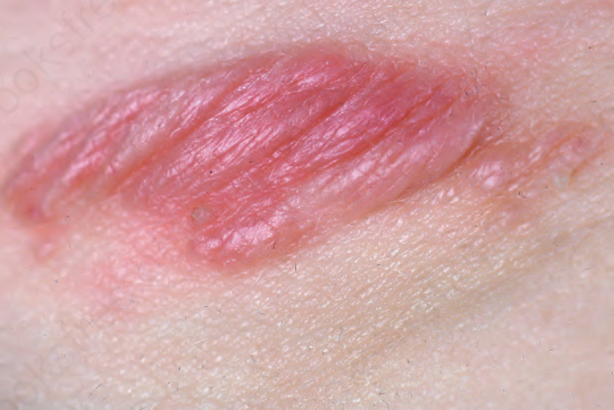
Fig. 29.347 Mastocytoma: edematous, erythematous plaque in a child. From the collection of the late N.P. Smith, MD, the Institute of Dermatology, London, UK.