Drug-induced pseudolymphoma
Drug-induced pseudolymphoma
This is discussed in Chapter 14.
The infiltrate consists of a mixed population of T and B cells.2,3 CD8+ T cells are sparse.3 In most cases, T cells predominate, but in some B cells are present in equal or greater numbers.4 The epithelioid histiocytes react with antibodies to CD68 whilst antibodies to S100 protein and CD1a highlight collections of perifollicular dendritic cells.2–4 There is no evidence of immunoglobulin light chain restriction in the B-cell rich variants.2–4
Gene rearrangement studies were uniformly negative in a Japanese series, but a recent large study from Europe demonstrated clonal T-cell receptor rearrangements by PCR in about 50% of patients, and clonal immunoglobulin gene rearrangement in three out of 42 cases.2,4 In one case, both the T-cell receptor and immunoglobulin heavy chain gene were clonally rearranged.4 Clonal cases of pseudolymphomatous folliculitis show no clinical or pathological differences to nonclonal ones.4 However, the median follow-up of 3 years in this study is too short to draw definite conclusions. If clonality is found, then close follow-up is advisable.4
1458 Cutaneous lymphoproliferative diseases and related disorders
It is questionable whether pseudolymphomatous folliculitis is truly distinct from other cutaneous lymphoid hyperplasias in which hair follicle hyperplasia is absent. However, it is important to distinguish pseudolymphomatous folliculitis from cases of lymphoma, especially as follicular hyperplasia has also been reported, albeit rarely, in a variety of primary and secondary cutaneous lymphomas of T- and B-cell type.4 The presence of solitary lesions, absence of an aberrant phenotype, and demonstration of polyclonality by light chain immunohistochemistry all favor a benign diagnosis. However, as evidenced above, care must be taken interpreting the results of gene rearrangement studies, as not all molecularly clonal lesions are lymphomas.
Atypical cutaneous lymphoproliferative disorder of HIV infection
Clinical features Also known as pseudo-Sézary syndrome, this rare condition presents with widespread, pruritic, and often erythematous patches, papules, and plaques.1–7 Lesions are sometimes lichenified.4,7 Hyperpigmentation is common and occasionally hypopigmentation is noted.5,7,8 Erythroderma, palmoplantar keratoderma, and photosensitivity are also described.2,5,6,8 Patients typically have advanced HIV disease with severe immunosuppression.5,7 Lymphadenopathy is present in a minority of patients, and circulating Sézary-like cells may be detected.2,8 The clinical course is chronic and may be modified by introduction of antiretroviral therapy.9 Exceptionally, cutaneous T-cell lymphoma supervenes.7
Pathogenesis and histologic features The etiology is unknown although the infiltrate has been shown to be HIV specific and directed toward a variety of antigens.8 There is no evidence of a drug-related pathogenesis.7 Photosensitivity may be an initiating event, at least in some patients.2 HTLV-I proviral DNA has been identified in the skin and peripheral blood of two patients but is absent in others.2,4
Histologically, there is a superficial and deep perivascular and perifollicular polymorphous infiltrate composed of lymphocytes, eosinophils, plasma cells, and rare neutrophils accompanied by an atypical mitotically active lymphoid population with enlarged irregular nuclei containing prominent nucleoli.2,7 Mycosis/Sézary cells may be present.2,4,7 Epidermotropism is variably present, being reported in some cases but described as absent or minimal in others.1,2–4,6–10
The lymphoid cells are CLA+, CD2+, CD3+, CD5+, CD8+, and TCRβ+.2,3,5–8 CD7 may be diminished.3,7 CD4, CD15, and CD30 are negative.7 B cells are absent. TCR gene rearrangement studies are negative.2,3,6
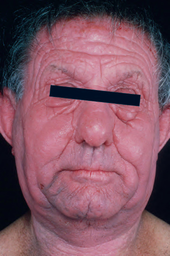
Fig. 29.180 Chronic actinic dermatitis: marked erythema, edema, and thickening of the skin. From the collection of the late N.P. Smith, MD, The Institute of Dermatology, London, UK.