Pustular mycosis fungoides
Pustular mycosis fungoides
Clinical features Pustular lesions are very rare in cutaneous T-cell lymphomas including MF and erythrodermic variants such that there can be confusion with pemphigus and subcorneal pustular dermatosis.1,2 A palmoplantar pustulosis-like variant has also been documented. Similar lesions may be encountered in patients with psoriasis and MF.3–5
1421 Classic mycosis fungoides with unusual clinical manifestations
Pathogenesis and histologic features The cause of pustulation is unknown. Histologically, the pustular lesions may be related to subcorneal neutrophil abscesses (i.e., true pustules) or else reflect conspicuous Pautrier microabscesses (Fig. 29.55).1,2
Differential diagnosis PAS and Gram stains should be performed to exclude a secondary infection. Pustular lesions have been described in a patient with MF as a result of demodex folliculitis.6
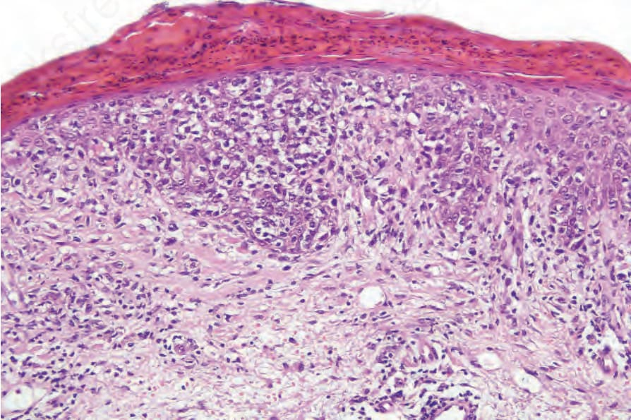
Fig. 29.55 Pustular mycosis fungoides: early pustule formation arising in a background of patch stage disease.
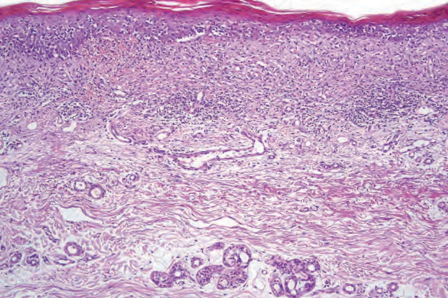
Fig. 29.56 Purpuric mycosis fungoides: this specimen comes from a patient with poikiloderma atrophicans vasculare.