Reticulohistiocytoma
Reticulohistiocytoma
Clinical features Reticulohistiocytoma is a rare benign conjunctival lesion that usually occurs as an isolated skin nodule or as part of multicentric reticulohistiocytosis, a systemic disorder. Reported cases of reticulohistiocytoma in the ocular
1384 Tumors of the conjunctiva
pigmented lesions, and endocrine overactivity.2 The lesion is a slow-growing, painless, well-circumscribed, yellow-pink, fleshy, translucent-to-solid mass on the bulbar conjunctiva.1,2
Histologic features Myxoma is a paucicellular tumor composed of spindle- and stellate-shaped cells within a loose mucoid, myxoid stroma.1–3 Some tumor cells may have small intranuclear vacuoles, and some may have clear intracytoplasmic inclusions. The stroma is composed of Alcian-blue-positive, hyaluronidase-sensitive hyaluronic acid mucopolysaccharides and chondroitin sulfates with a network of reticulin fibers, small blood vessels, and collagen fibers. Few mast cells may be present in the stroma.2,4 Histochemical stains for mucicarmine and colloidal iron are also positive in the tumor stroma, while PAS and oil red O stains are negative. By immunohistochemistry, tumor cells show strong immunoreactivity for vimentin and focal reactivity for a-smooth muscle actin, but negative for S100 protein, desmin, and myoglobin.1,5 Ki-67 proliferation index is low (< 5% of tumor cells).4
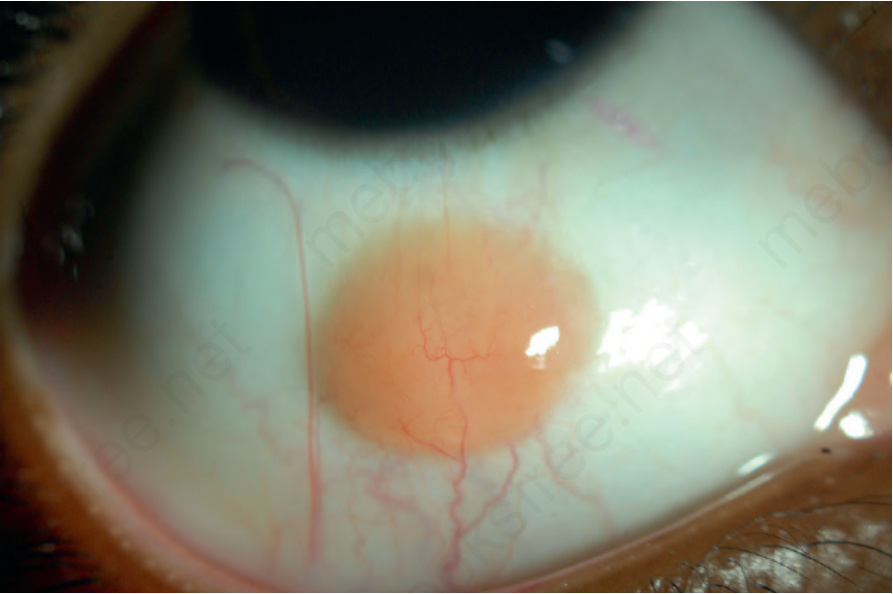
Fig. 27.53 Juvenile xanthogranuloma: a circumscribed yellow-orange lesion is on the inferior bulbar conjunctiva.