Spitz nevus
Spitz nevus
Clinical features Conjunctival Spitz nevus is a very rare melanocytic lesion that usually occurs in children and adolescents.1,2 The lesion is nonpigmented and grows rapidly, raising suspicion for melanoma, although melanoma is extremely uncommon in children.
Histologic features In contrast to typical conjunctival nevus, which may contain spindle cells within the epithelium that are oriented parallel to the surface, Spitz nevus
Clinical features Inflamed juvenile conjunctival nevus is a benign, usually amelanotic, juxtalimbal lesion in children and adolescents (Fig. 27.32). It may grow rapidly, raising concern for malignancy. Patients often have history of allergy, allergic conjunctivitis, or vernal conjunctivitis.1 This association is supported by one study that showed increased expression of nerve growth factor, eosinophils, and mast cells in inflamed juvenile nevus and modulation of eosinophil properties by lesional fibroblasts, partly through nerve growth factor.2 Intralesional cysts may be seen clinically.
Histologic features Inflamed juvenile conjunctival nevi have intraepithelial and stromal melanocytic nests with solid and cystic inclusions, goblet cells, and chronic inflammatory infiltrates with eosinophils and plasma cells (Fig. 27.33).1,3 Rapid growth of a clinically inflamed nevus indicates inflammatory infiltration and cystic enlargement rather than malignant transformation.
1375 Melanocytic tumors
Congenital melanosis oculi (congenital ocular melanocytosis)
Clinical features Congenital melanosis oculi or congenital ocular melanocytosis is characterized by flat, spiculated slate-gray pigmentation of the sclera sometimes surrounding episcleral lymphatics and blood vessels (Fig. 27.34). The periocular skin, orbit, meninges, and soft palate may be affected.1 Although the conjunctiva is not involved, the lesion is included here because it is considered in the clinical differential diagnosis of conjunctival pigmented lesions. When periocular skin is involved, the condition is called nevus of Ota or oculodermal melanocytosis. Patients with congenital melanosis oculi have approximately 1 in 400 risk of developing uveal melanoma; however, conjunctival melanoma has not been described in these patients.2
with dark skin complexion, including African-Americans, Hispanics, Asians, and whites.1 The pigmentation is concentrated around the limbus and may involve the cornea. It rarely involves the fornix or palpebral conjunctiva. The distribution of pigmentation may be asymmetric, but this should raise suspicion for PAM, a unilateral, potentially premalignant condition (see next section).
Histologic features Pigmented dendritic melanocytes are present in the episcleral and scleral tissues (Fig. 27.35). The conjunctiva is not involved.
Complexion-associated conjunctival pigmentation (racial melanosis)
Clinical features Complexion-associated melanosis is a common, bilateral condition characterized by diffuse or patchy, flat pigmentation of the conjunctiva in patients
Histologic features Histologically, there is hyperpigmentation of basal conjunctiva squamous epithelial cells without melanocytic hyperplasia or cytological atypia.1
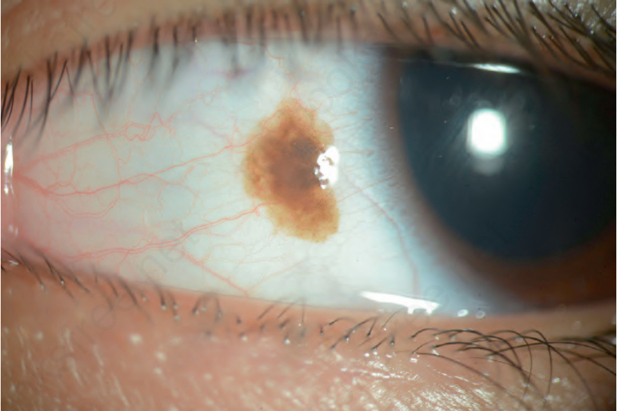
Fig. 27.32 Inflamed juvenile nevus: partially pigmented inflamed juvenile nevus on the bulbar conjunctiva at 9 o’clock. Intralesional cysts are seen.
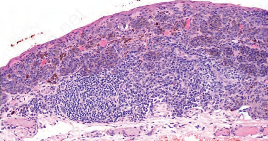
Fig. 27.33 Inflamed juvenile nevus: nests of nevus cells are admixed with lymphocytes and plasma cells.
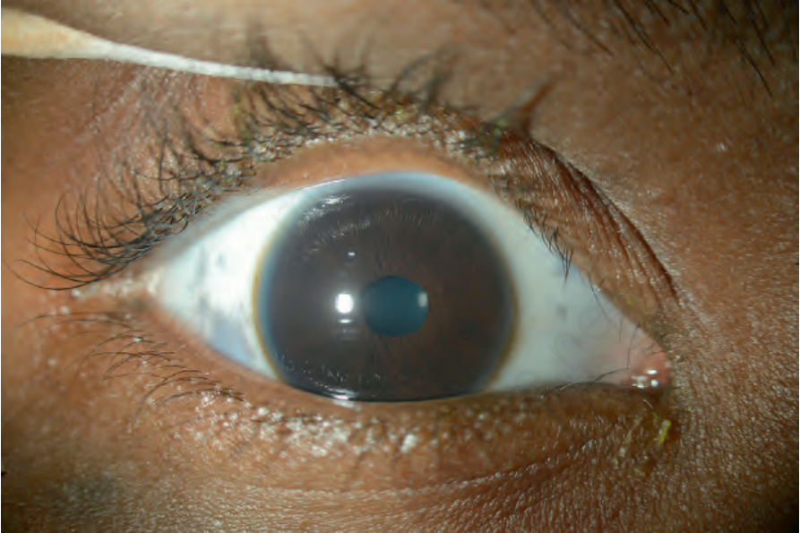
Fig. 27.34 Congenital melanosis oculi: clinical view of the right eye showing slate gray pigmentation of the sclera.
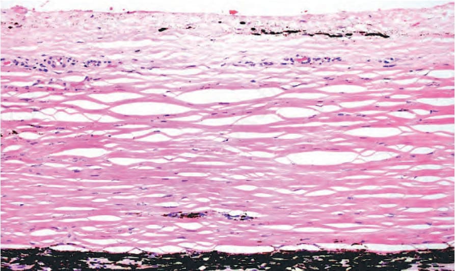
Fig. 27.35 Congenital melanosis oculi: pigmented dendritic melanocytes are present within episcleral and scleral tissues. The choroid is also heavily pigmented.