Meyerson nevus
Meyerson nevus
Clinical features Meyerson nevus represents an annular dermatitis (eczema) superimposed upon an acquired benign melanocytic nevus, usually a compound nevus, and is of unknown etiology (Fig. 25.105).1–4 This phenomenon has also been reported in congenital melanocytic nevi, dysplastic nevi, common acquired nevi, Spitz nevi, melanoma in situ, and in invasive melanoma.5–9
Meyerson nevus presents with a sometimes pruritic, erythematous, scaly border measuring up to 1.0 cm in width.2 Lesions, which are often multiple, appear most often on the trunk and proximal extremities, show a male predominance, and are most commonly seen in the third decade. Following resolution of the dermatitis, the nevus appears unchanged.1,2 The evolution of a Meyerson nevus into a halo nevus and coexistence of the two types of nevi in the same patient have been reported.10,11
Histologic features The histologic features are those of parakeratosis, acanthosis, and spongiosis associated with a superficial perivascular chronic inflammatory cell infiltrate (Figs 25.106–25.108). The spongiotic process is occasionally so prominent that the background melanocytic proliferation is obscured and may be missed.9 In addition, spongiosis can be associated with significant distortion of the morphology of intraepidermal melanocytes, including the appearance of more epithelioid melanocytes and limited upward migration of tumor cells.9 This is usually associated with mild cytological atypia.9
The inflammatory cell infiltrate consists predominantly of CD4+ lymphocytes.5,12 Eosinophils are often seen and may be conspicuous, and
eosinophilic spongiosis is sometimes identified.2 There is no histologic evidence of regression.2
Similar changes surrounding a wide range of lesions, including seborrheic keratosis, stucco keratoses, keloid, nevus flammeus, and squamous cell carcinoma, have rarely been described.13,14
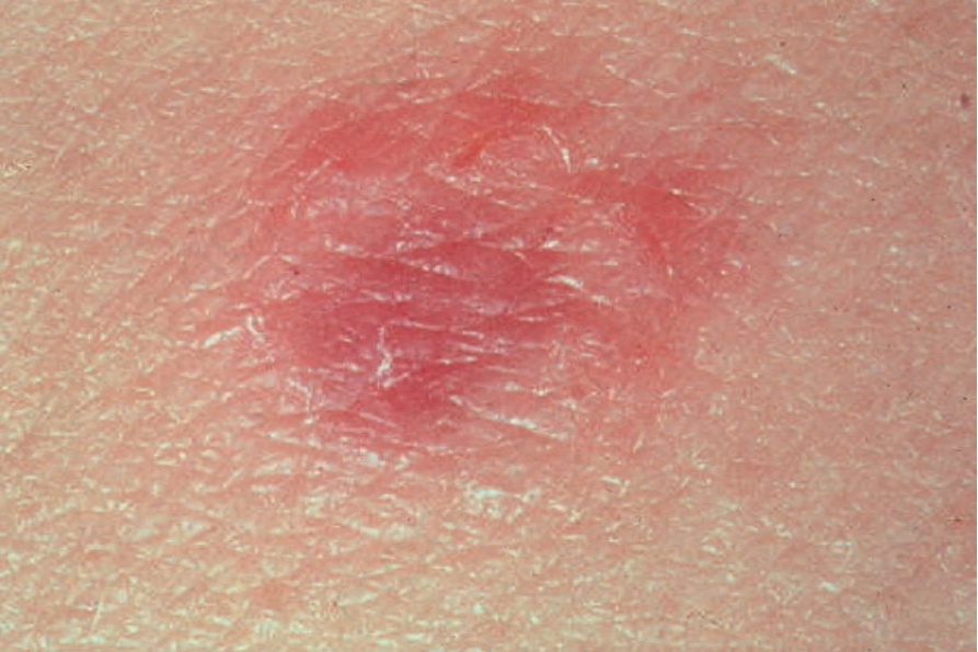
Fig. 25.105 Meyerson nevus: there is intense erythema surrounding this banal dermal nevus. By courtesy of the Institute of Dermatology, London, UK.
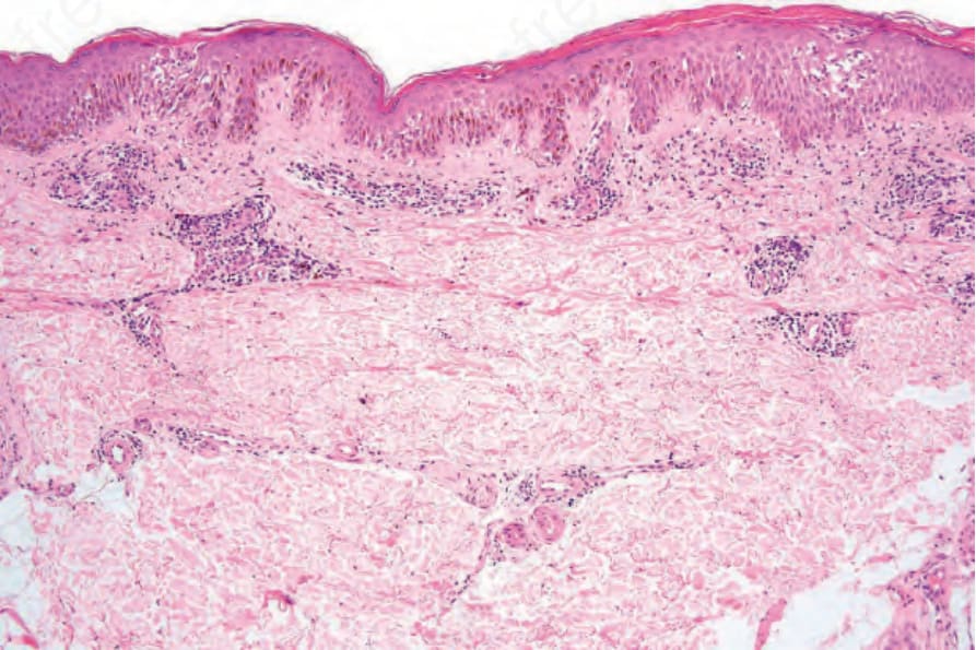
Fig. 25.106 Meyerson nevus: several junctional nests are evident in a background of spongiotic dermatitis.
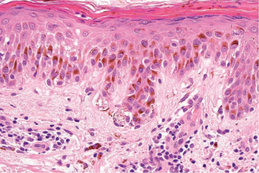
Fig. 25.107 Meyerson nevus: high-power view of Fig. 25.106.
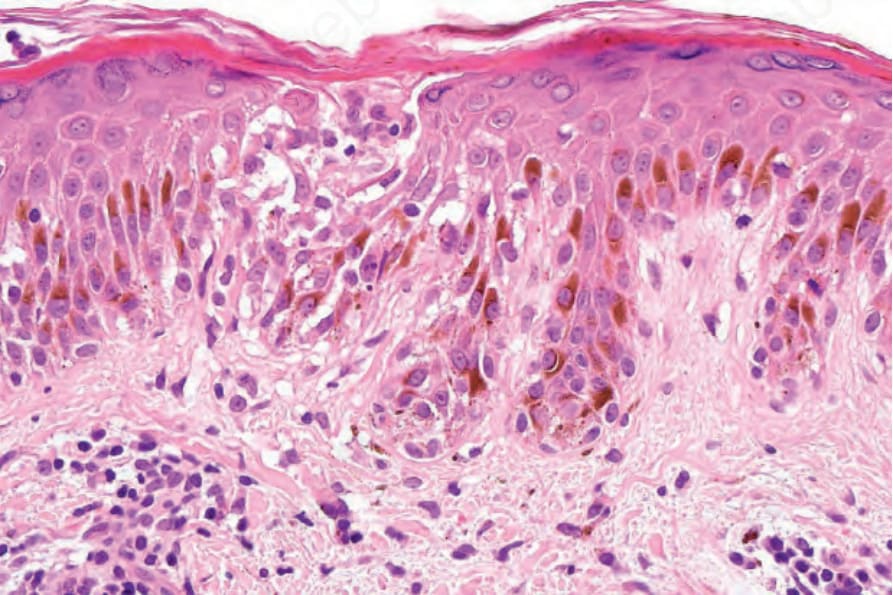
Fig. 25.108 Meyerson nevus: there is spongiosis with a microvesicle.