Nevi of flexural skin
Nevi of flexural skin
Flexural sites are defined as sites with cutaneous folds, including axilla, mammary folds, popliteal and antecubital fossae, umbilicus, pubis, scrotum, and perianal skin, but also folds on the neck and abdomen.1,2 Nevi at flexural sites have predilection for umbilicus and axilla, show equal gender distribution, and have an average size of less than 1 cm. They are well circumscribed and symmetrical.1 Interestingly, development of agminated flexural melanocytic nevi, involving most commonly the inguinal area and axilla, has been reported in children with a history of Langerhans cell histiocytosis.3–5
Histologic features The peculiar histologic features of nevi at flexural sites include enlarged junctional nests, variation in the size and shape of nests, confluence of
1254 Melanocytic nevi
nests, and diminished cohesion of melanocytes within the nests – a so-called nested and dyshesive pattern of melanocytic proliferation.1 The nests are localized at the tips and sides of the rete ridges. Involvement of skin adnexa is not uncommon. Some degree of nuclear atypia is invariably present. Focal fibrosis at the tips of the rete ridges, often of the lamellar type, can be seen.
A further subset of melanocytic nevi in the umbilicus is characterized by more extensive lentiginous proliferation of melanocytes displaying moderate degree of cytological atypia, focal and limited upward migration, as well as prominent lamellar fibrosis, often extending into the reticular dermis.6 This peculiar type of lamellar fibrosis frequently contains entrapped melanocytic nests displaying mild cytological atypia, with either horizontal or irregular arrangement of the collagen bundles.6
The dermal melanocytic component is unremarkable, maturation is retained, and dermal mitoses are usually absent.
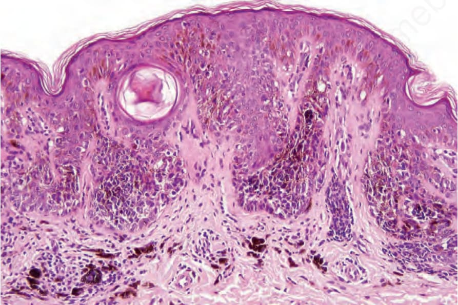
Fig. 25.77 Nevus of breast: the nests are large and dyscohesive.
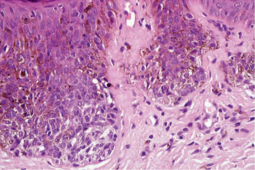
Fig. 25.78 Nevus of breast: scattered severely atypical nevus cells are present with large vesicular nuclei and prominent nucleoli. There is also a conspicuous dendritic cell population.
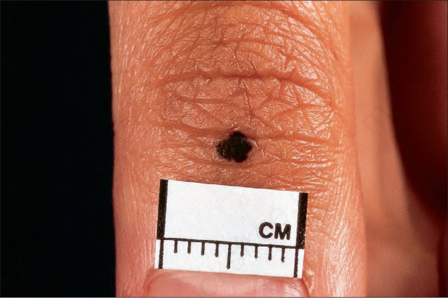
Fig. 25.79 Acral nevus: note the intense pigmentation and irregular border. By courtesy of the Institute of Dermatology, London, UK.