Lichen striatus
Lichen striatus
Clinical features Nail lichen striatus is rare but probably under-reported. It has mainly been described in children and young adults and usually affects a single digit. It is characterized by a median or asymmetrical, longitudinal, lichenoid nail plate dystrophy (Fig. 23.30).1 Upper limbs are more frequently affected than the lower limbs. When the typical cutaneous involvement characterized by small, flesh-colored papules in a linear distribution is present, diagnosis is easy. However, the nail dystrophy may appear before the skin involvement2 or can be isolated. Spontaneous regression after a median duration of 22.6 months is the rule.1
Differential diagnosis Multinucleate giant cells in the nail bed epithelium were thought to be specific to Darier disease. In fact, they may also be observed in several unrelated nail conditions such as onychopapilloma4,5 (see erythronychia) and Bowen disease. A case of focal subungual warty dyskeratoma and three cases of acantholytic dyskeratotic acanthomas of the nail have been described.6,7 Clinically, they presented as median longitudinal erythronychia with distal onycholysis. Histologically, they were characterized by suprabasal clefts, acantholytic cells, grains and corps ronds.
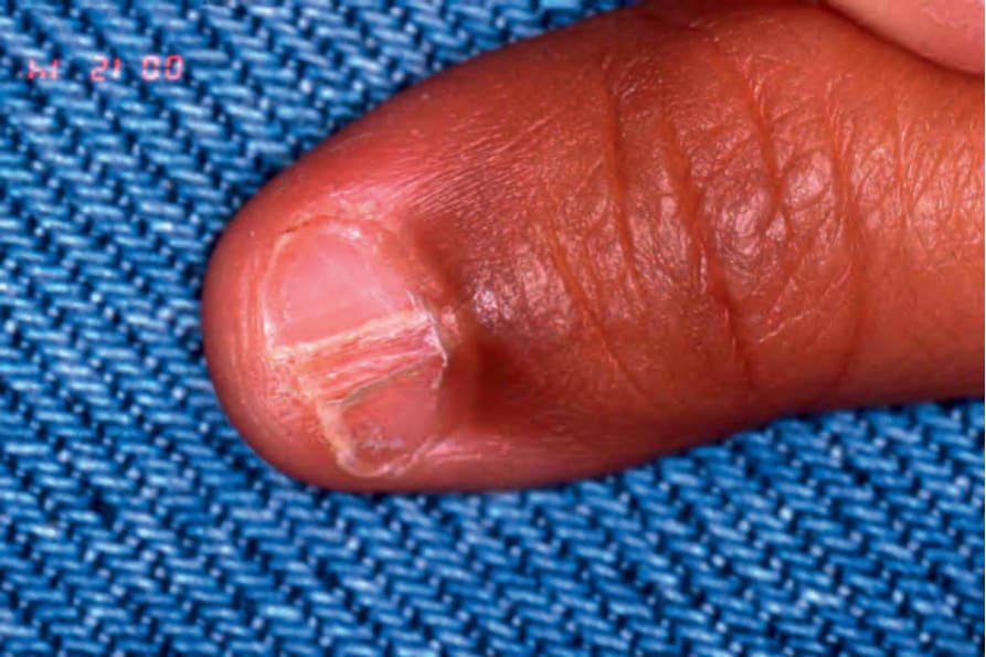
Fig. 23.30 Lichen striatus: there is a median, longitudinal, lichenoid nail involvement. Discrete flesh-colored linear papules are present on the dorsum of the finger. The patient is a 2-year-old boy.