Trichoschisis and trichoclasis
Trichoschisis and trichoclasis
Trichoschisis means a clean transverse fracture across the hair shaft involving both the cuticle and the cortex (Fig. 22.176).1 It develops as a consequence of loss of cuticular cells and is frequently associated with TTD.2,3 It has also been observed in patients with normal hair.4
Trichoclasis refers to a greenstick fracture of the hair shaft. It is therefore an oblique or transverse incomplete fracture involving the cortex but
1113 Fractures of the hair shaft
characterized by a generalized desquamative erythroderma. The defects of the hair shaft appear early in life and involve all the hairs in the body. In a minority of patients the manifestations are very mild. In typical cases, however, the hair is short, dry, dull, and breakable. Eyelashes and eyebrows are scarce or absent. The eyebrows are more commonly and severely affected than the scalp and therefore represent the site of choice for a biopsy.2 Although Netherton syndrome has been associated with aminoaciduria, this is not a constant finding.
Other clinical features that have been reported are neurological defects, mental retardation, short stature, delayed growth, recurrent infections, and hypo- or hypergammaglobulinemia.3,4
Pathogenesis and histologic features Netherton syndrome is caused by a mutation in the SPINK5 gene at chromosome 5q32, which encodes an inhibitor of serine protease called LEKT1 (lymphoepithelial Kazal-type related inhibitor), a new type of serine protease inhibitor involved in the regulation of skin barrier formation and immunity. It is thought that the alteration in the epidermal expression of LEKT1 leads to the premature activation of proteolytic enzymes in the stratum corneum with separation of corneocytes.5–7 SPINK5 testing can be performed as an aid in the diagnosis of Netherton syndrome by immunohistochemistry of a skin biopsy with an antibody against LETK1.8
leaving the cuticle intact.1 The sulfur content in the cuticle and the cortex is normal. It is not a specific sign and can be seen associated with a wide range of hair shaft disorders (Fig. 22.177).
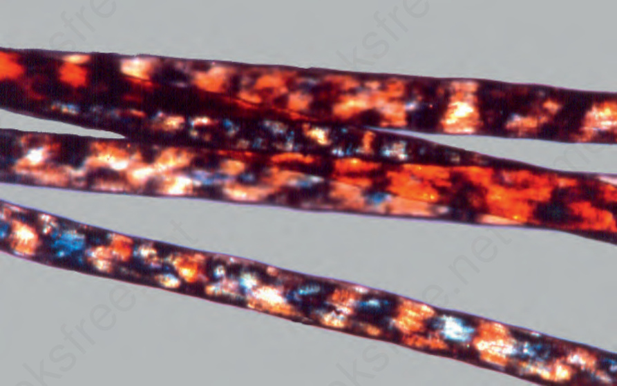
Fig. 22.175 Trichothiodystrophy: typical tiger tail appearance in hair shafts under polarized light. Courtesy of P. Reygagne, MD, Centre Sabouraud, Paris, France.
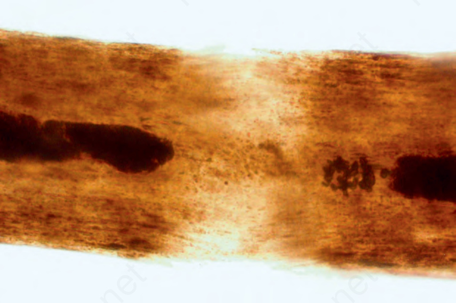
Fig. 22.176 Trichoschisis: note a crack in the cuticle that is perpendicular to the axis of the hair fiber; the cortical fibers do not protrude.
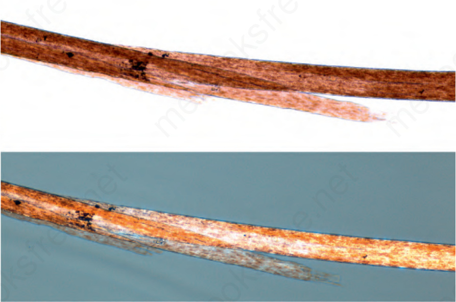
Fig. 22.177 Trichoclasis ‘Greenstick’ fracture of the hair shaft, consisting of an oblique fracture splinted by an intact cuticle, easily visible with polarized light (bottom).