Embryology
Embryology
Embryologically, each follicle is composed of epithelial and mesenchymal components. Epithelial invaginations (placodes) derive from the fetal epidermis and project downward at regularly spaced intervals, proliferating under the influence of the underlying connective tissue cells which form the mesenchymal condensation of the hair peg. This occurs around the 10th week of gestation (Fig. 22.10). This dermal condensate will eventually mature into the dermal papilla. The hair follicle does not develop in the absence of this mesenchymal influence. Afterwards, the hair follicle elongates into a lace of epithelial cells and the deeper component forms the bulbous and the matrix cells, which give origin to the hair shaft and inner root sheath. The outer root sheath forms three bulges. The upper one will give origin, in the follicles located in the anogenital region, axillae, areolae, periumbilical region, eyelids, and external ear canals, to the apocrine glands. The middle bulge will give origin to the sebaceous gland. The lowermost bulge corresponds to the location of the epithelial stem cells. This is also the attachment site of the arrector pili muscle.1
of the newborn and vellus hairs on the rest of the body. No new hair follicles form after birth. At puberty the vellus hairs present on genital skin and axilla in females and on the legs, torso, and the chin in males transform into terminal hairs under the influence of sexual hormones. Paradoxically, in individuals with androgenic alopecia the terminal hairs in the scalp transform into miniaturized hair follicles in those individuals who are susceptible. Hair follicles will eventually be found on all body surfaces except for the palms, soles, and mucous membranes.
The genes and molecules that participate in the development of the hair follicle have been extensively studied. Different positive and negative regulators which are expressed at variable time during the development of hair follicles have been identified.2,3 Some of the most relevant of these regulators are described in Table 22.3.
The first follicles develop in the scalp, and from there they extend downwards to populate the rest of the body. Initially, they are observed as fine thin nonpigmented hairs, known as lanugo hair. These fall off at the end of the gestation period, although some remain until after birth. The majority are replaced by terminal hairs, present in the scalp, eyelashes, and eyebrows
Box 22.1 Hair biopsy report: basic information to be evaluated and included in a histology report of a 4.0 mm punch biopsy.
Total number of hair follicles in the biopsy (terminal + vellus, in anagen,
catagen, and telogen) Total number of hair follicles per square millimeter (total/12.6) Number of terminal hair follicles Number of vellus hairs (vellus + miniaturized hairs) Number of undetermined follicles Terminal:vellus hair ratio (T:V): terminal hair follicles / vellus Number of terminal hair follicles in anagen Number of terminal hair follicles in telogen Number of terminal hair follicles in catagen Telogen count: terminal telogen hairs (telogen + catagen) / terminal hair
follicles. Presence, type and localization of inflammatory cell infiltrate Presence or absence of scar tissue Presence or absence of pigmented casts
Characteristic Asia America
Race Taiwanese, n = 31 Korean, n = 35 Iranian, n = 30 Caucasian, n = 22 African American, n = 22
Author Present study Lee et al5 Aslani8 Whiting2 Sperling3 p-Value*
Age, mean ± SD 37.0 ± 15.3 33.1 ± 10.0 35.5 ± 14.7 43 ± 3.5 31.7 ± 8.5 .04
Number of terminal hairs, mean ± SD 20.5 ± 4.6 14.9 ± 3.2 34 ± 6.4 35 ± 2.1 18.4 ± 5.0 <.001
Number of vellus hairs, mean ± SD 0.8 ± 1.0 1.1 ± 1.3 2.4 ± 1.2 5 ± 0.6 3.0 ± 2.1 <.001
Number of total hairs, mean ± SD 21.3 ± 4.8 16.1 ± 3.6 36.3 ± 7.2 40 ± 2.2 21.5 ± 5.0 <.001
Number of follicular units, mean ± SD 9.4 ± 1.9 7.8 ± 1.7 ND 14 ± 0.5 ND <.001
Anagen: telogen ratio,% 91.6 : 8.4 93.6 : 6.4 93.7 : 6.3 93.5 : 6.5 93.9 : 6.1 .47
Terminal: vellus ratio 25.3 : 1 13.5 : 1 17.4 : 1 7 : 1 6.1 : 1 <.001
Density of hair follicles/mm2 1.69 ± 0.4 1.2 ± 0.3 2.89 ± 0.6 3.1 ± 0.8 1.65 ± 0.4 <.001
Density of follicular units/mm2 0.75 ± 0.2 0.62 ± 0.1 ND 1.11 ± 0.04 ND <.001
*Analysis of variance. SD, standard deviation; ND, no data. *Ko J.H., Huang Y.H., Kuo T.T. Hair counts from normal scalp biopsy in Taiwan. Dermatol Surg. 2012. 38, Sep;1516-20.
Hair Follicle Morphogenesis Step Gene Product
Outer root sheath Sox9, Shh/Smo
Inner root sheath GATA3, Cutl1 (CDP), BMPs/BMPR1a, Shh/ Smo, Notch1/Jagged/ Delta/RBP-Jk for IRS fate maintenance
1057 Embryology and anatomy of the normal hair follicle
Hair shaft B-catenin, Wnt’s and Lef-1, VDR (Vitamin D receptor)(only in postnatal hair cycle), BMPs/ BMPR1a, Shh/Smo, msx1 and 2, FoxN1/Nude, HoxC13, Notch1/Jagged/Delta/RBP-Jk (only in postnatal hair cycle)
Polarity, shaping and bending
Shh (asymmetric, polarized expression pattern in hair matrix), Igfbp5, Eda A1, Krox-20, FoxE1, Runx3, Sox18
Innervation B-catenin, NCAM
Pigmentation FoxN1/Nude, SCF/c-kit, Notch
Bulge region/hair stem cell maintenance
BMPs/BMPR1a, Lhx2, Sox9, NFATc1, p63, Tcf3 (w/o Wnt activity), Rac1, integrins a3b1 and a6b4
Note: Transcription factors in Italics Modified from Schneider MR, Schmidt-Ullrich R, Paus R. The hair follicle as a dynamic miniorgan. Curr Biol. 19, 2009;R132-42.
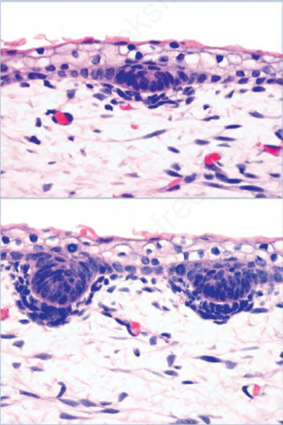
Fig. 22.10 Fetal hair follicle, upper and lower panel: epidermal precursor and primary hair germ with underlying connective tissue cells (dermal condensate).
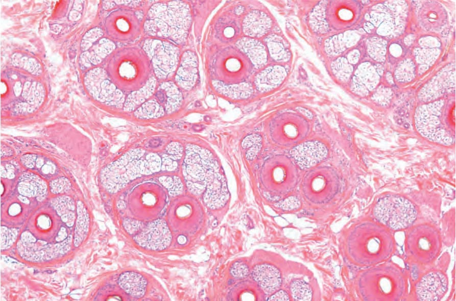
Fig. 22.11 Hair biopsy, horizontal section: the follicular units are clearly delineated by condensation of the adventitial collagen (Masson trichrome stain).
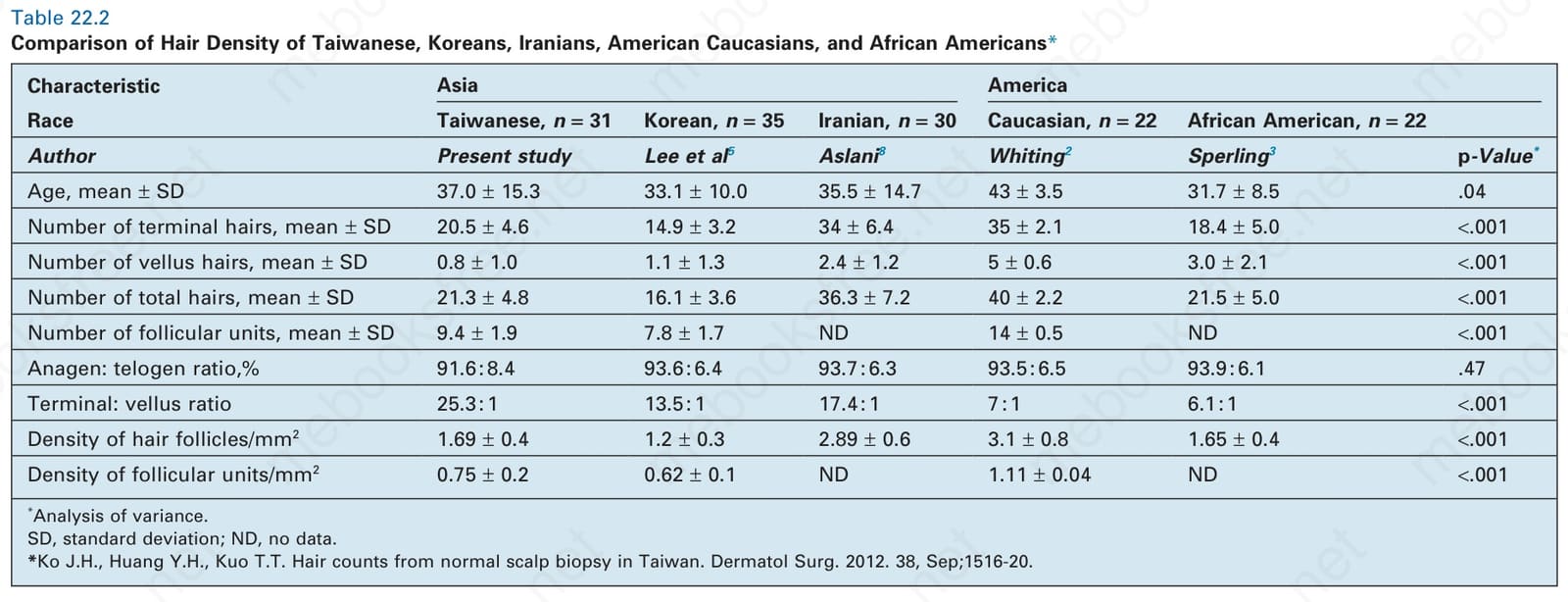
Table 22.2 Comparison of Hair Density of Taiwanese, Koreans, Iranians, American Caucasians, and African Americans*
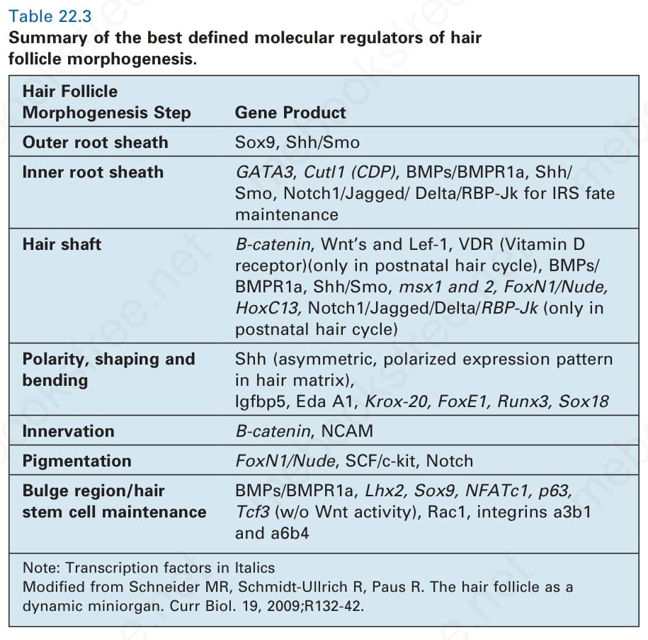
Table 22.3 Summary of the best defined molecular regulators of hair follicle morphogenesis.