Anetoderma
Anetoderma
Clinical features Anetoderma (Gr. anetos, slack) is an acquired, purely cutaneous disorder characterized by numerous 0.5–3.0-cm foci of atrophic or wrinkled, flaccid skin, due to loss of elastic fibers, which may be accompanied by herniation into the subcutaneous fat (Figs 21.59 and 21.60).1,2 Lesions, which are most common in young adults, particularly affect the upper trunk and proximal arms, although they may be more widespread.3 One case was reported to have a granuloma annulare-like pattern.4 An unusual linear form has been described.5 There is a female predominance, and most patients range in age from 20 to 40 but all ages are affected.2,3,6,7
PXE-like changes are sometimes described in the setting of vitamin K-dependent factor deficiencies and hemochromatosis. The association with vitamin K-dependent factor deficiencies lends credence to the theory that vitamin K cofactor may be involved in inherited pseudoxanthoma elasticum.24,26–29 Of note, inherited pseudoxanthoma elasticum is made worse by warfarin use, which affects the vitamin K-dependent synthesis of calcium-dependent clotting factors, as demonstrated by surveying a subset of the 4000 patients in the PXE International database.30 A mouse model confirmed this association.30
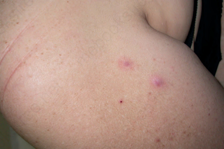
Fig. 21.59 Primary anetoderma: atrophic lesions with a wrinkled appearance. Courtesy of J.C. Pascual, MD, Alicante, Spain.