Pinta
Pinta
Clinical features This is a nonsexually transmitted treponematosis characterized by depigmented skin lesions. It is caused by Treponema carateum (T. pallidum subsp. carateum), an organism that is very similar to T. pallidum subsp. pallidum.1–5 It is generally confined to remote regions in tropical Central and South America where the inhabitants live in poor hygiene and in close proximity.1,3,5–7 The condition may nevertheless be encountered in nonendemic countries among migrants and refugees from endemic areas.8 Children, adolescents, and young adults are primarily affected, and transmission is thought to be by direct cutaneous or mucous membrane contact, possibly via minute abrasions.1–3,6,7,9
The lesions present as small scaly erythematous indurated papules and plaques on exposed skin, usually on the hands and feet. These disappear, but recur in a more disseminated form (pintids).1,3,6,7 Regional lymphadenopathy may occur.3 Late stages of pinta are characterized by disfiguring hyperpigmentation, achromia, hyperkeratosis, and atrophy (Fig. 18.145).1,3,7,10
B
Unlike syphilis and yaws, all manifestations of the infection are limited to the skin, and there is no evidence of systemic disease.3
Histologic features Histologically, early primary and secondary pinta lesions show hyperkeratosis and acanthosis, while later lesions demonstrate epidermal atrophy and a diminished basal melanin pigment concentration.1,5,10–13 Lymphocytic exocytosis, mild spongiosis, basal cell hydropic degeneration, and pigmentary
880 Infectious diseases of the skin
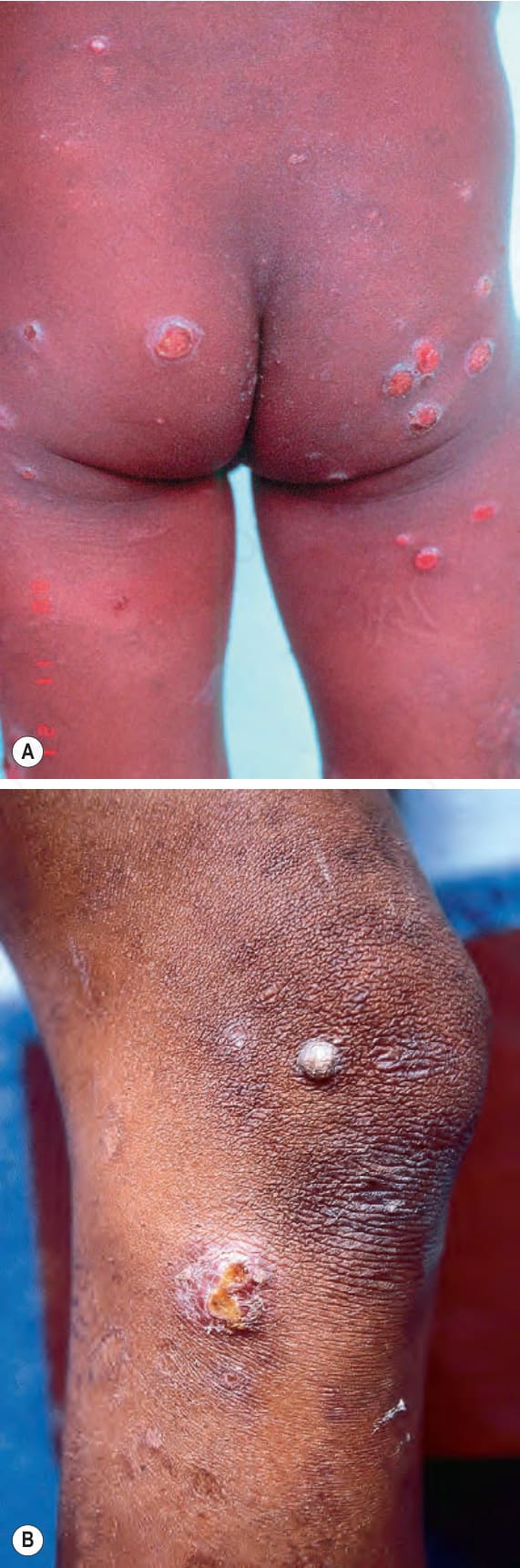
Fig. 18.140 (A, B) Early yaws: multiple smaller ‘daughter yaws’ may be widely distributed and usually present 2–4 months after the ‘mother yaw’. By courtesy of H.J.H. Engelkens, MD, and E. Stolz, MD, University Hospital, Rotterdam-Dijkzigt and Erasmus University, Rotterdam, The Netherlands.
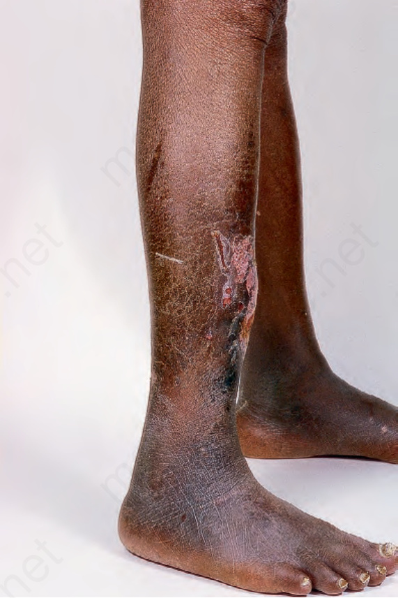
Fig. 18.141 Late yaws: note the bowing of the lower leg with cutaneous ulcerated and crusted lesions in this late stage. By courtesy of R.A. Marsden, MD, St George’s Hospital, London, UK.
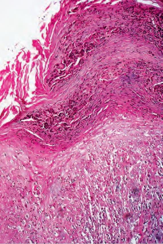
Fig. 18.143 Early yaws: biopsy through an evolving papilloma. There is very marked parakeratosis associated with abundant neutrophil debris. The epidermis shows intense acute inflammation. By courtesy of H.J.H. Engelkens, MD, and E. Stolz, MD, University Hospital, Rotterdam-Dijkzigt and Erasmus University, Rotterdam, The Netherlands.
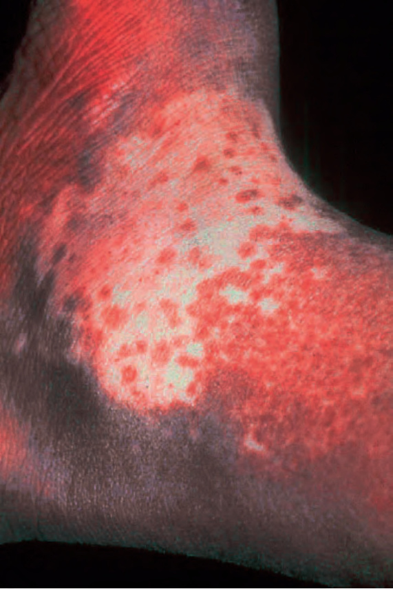
Fig. 18.145 Pinta: this is a late lesion showing characteristic complete loss of pigmentation surrounded by a hyperpigmented border. By courtesy of R. Arenas, MD, and J. Salas, MD, Azteca, Monterrey, Mexico.