Trench fever
Trench fever
Clinical features Although a major epidemic of trench fever was documented during the First World War, more recent outbreaks of this condition have been described among homeless people, in whom there is a high seroprevalence of this bacteremic illness.1,2 Outbreaks have also occurred in overpopulated Central African refugee camps.3 Trench fever is caused by Bartonella quintana; human body lice (Pediculus humanus var. corporis) are the known vectors.1,2,4 Head lice (P. humanus var. capitis) might also play a role in the transmission of the disease.5,6 B. quintana may also cause chronic bacteremia, endocarditis, and BA (see below).4,7
severe hemolytic anemia. The last is attributable to infection of the circulating erythrocytes and can be confirmed with the aid of a blood smear. The case fatality rate among untreated patients may exceed 80% during acute outbreaks. Later, the disease enters an eruptive phase characterized by the evolution of numerous papular, nodular, or verrucous vascular skin lesions, referred to as verruga peruana (Peruvian wart, cutaneous verrucous disease).2,4–6 These occur predominantly on the face and extremities (Fig. 18.128). Atypical cases may present with verrucous skin lesions as the sole manifestation.2 Most lesions resolve spontaneously.3 Genital lesions and nasal mucosal involvement have been recorded.7
The condition is endemic in the higher altitude regions of Peru, where it was first described in the nineteenth century. Carrión disease also occurs in Ecuador and Colombia.5,8,9 Outbreaks have also been recorded in nonendemic parts of Peru.10 It has been suggested that the condition may be underreported in some endemic areas because of the existence of mild infection by less virulent strains of B. bacilliformis.2,10 The sand fly, Lutzomyia (Phlebotomus) verrucarum is the apparent vector.8
Patients present with non-specific symptoms and signs including headache, malaise, pyrexia, rigors, tachycardia, myalgia, arthralgia, and injected conjunctivae. An erythematous macular or papular skin rash may occur. The rash is often seen on the trunk and usually lasts no more than a day or two.8 The disease is rarely fatal, except in some debilitated patients. Relapsing illness sometimes occurs, and the organism may remain latent in the host for a number of years following the acute infection.8
Pathogenesis and histologic features There is infection of endothelial cells and circulating erythrocytes following introduction of B. bacilliformis via the bite of the vector. In the cutaneous verruga peruana lesions, organisms are detectable in the extracellular spaces, where they induce angiogenesis by producing putative microbial-encoded or microbial-induced angiogenic factors.1,11 In vitro studies have shown that B. bacilliformis exercises a mitogenic effect on human vascular endothelial cells. GroEL produced by the organism regulates endothelial cell growth.12,13 Others have observed that infection leads to the production of angiopoietin-2 by endothelial cells, and the production of VEGF by epidermal cells.14 The dermal angiomatous proliferation appears to occur in concert with the reactivation of latent B. bacilliformis organisms.1
Histologic features The histopathological features in the skin are non-specific. There is a perivascular lymphocytic infiltrate, without evidence of vascular thrombosis.8 Organisms are not usually demonstrable in routinely stained skin biopsy specimens. Dermal vascular proliferation and neutrophilic infiltration (as seen in BA) are not features of trench fever. The diagnosis is confirmed by serology, culture, or PCR.9
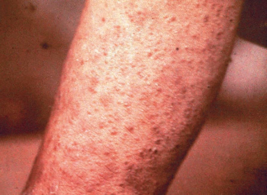
Fig. 18.128 Verruga peruana: widespread papules are present. By courtesy of F. von Lichtenberg, MD, Brigham and Women’s Hospital and Harvard Medical School, Boston, USA.