Secondary cutaneous mucinoses
Secondary cutaneous mucinoses
Clinical features Mucinous lesions on the skin have been described as part of a reactive process associated with various triggers. A patient recently developed multiple erythematous to skin-colored papules following infection with varicella-zoster virus.1,2 The lesions occurred in the same dermatome affected by postherpetic neuralgia and resolved as the pain improved. Mucinosis complicated by cutaneous necrosis can occur following injection with interferon. Recombinant interferon-beta-1b and interferon alfacon-1 have been implicated in the setting of treatment for multiple sclerosis and hepatitis C, respectively.3,4 Erythematous ulcerative plaques develop at injection sites. Additionally, mucinosis papules and plaques have been described in the vicinity of a recently replaced joint.5
Histologic features These mucinous disorders are characterized by intradermal mucin without a significant increase in dermal fibroblasts.1–5 Ulcerative lesions associated with interferon injections can also demonstrate intravascular thrombi.4
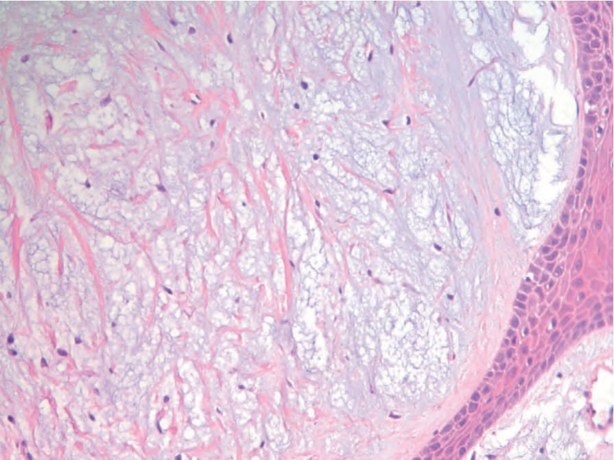
Fig. 13.192 Hereditary progressive mucinous histiocytosis. Nodular dermal infiltrate of histiocytes in a mucinous/myxoid background. By courtesy of C-K Sui, S-C Chao and J. Y-Y Lee, Tainan, Taiwan.