Myxoid cyst
Myxoid cyst
Clinical features Cutaneous myxoid cyst, sometimes inappropriately referred to as synovial cyst, presents as a soft or fluctuant cystic nodule on the dorsal aspect of the distal interphalangeal, the metacarpophalangeal and, less frequently, the metatarsophalangeal joints (Figs 13.188 and 13.189).1,2 An exceptional
624 Degenerative and metabolic diseases
A
B
(Fig. 13.190). The overlying epidermis is often atrophic and hyperkeratotic, and acanthosis may be seen at the edges. Early lesions are sometimes indistinguishable from cutaneous focal mucinosis. There is no evidence of any connection with an underlying joint.
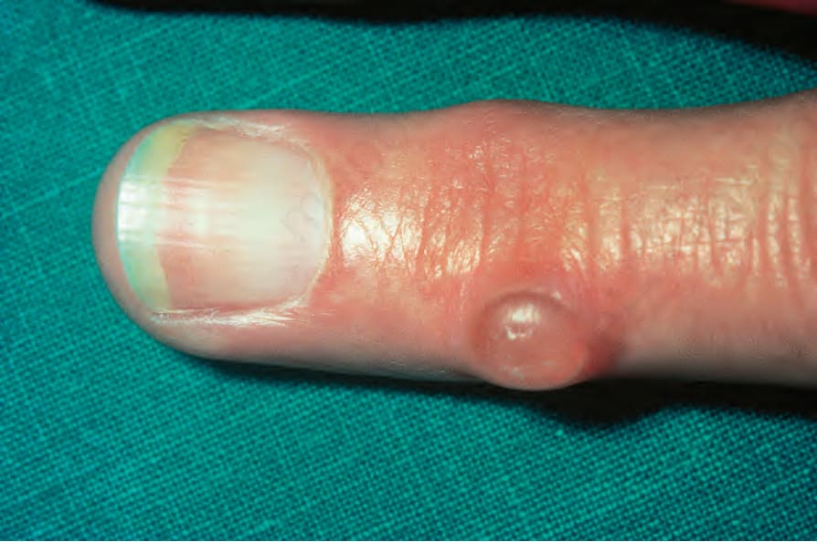
Fig. 13.188 Myxoid cyst: the translucency is typical. By courtesy of R.A. Marsden, MD, St George’s Hospital, London, UK.
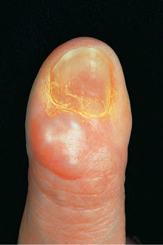
Fig. 13.189 Myxoid cyst: localization over the distal interphalangeal joint is characteristic. By courtesy of the Institute of Dermatology, London, UK.
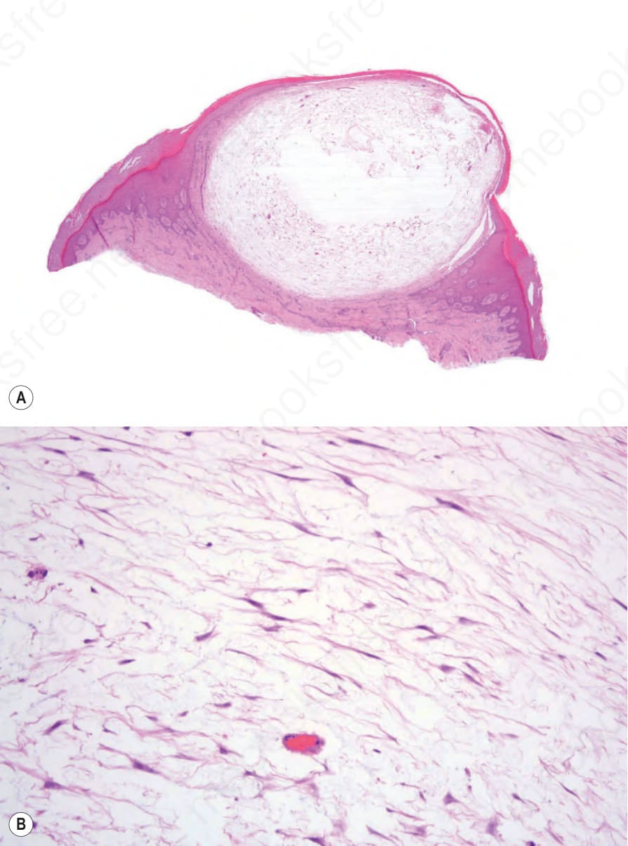
Fig. 13.190 (A, B) Myxoid cyst: excessive mucin deposition has resulted in this fluidfilled cyst. The overlying epithelium may appear attenuated or verrucous and occasionally the cyst is, in part, intraepidermal in location.