Periurethral cyst
Periurethral cyst
Clinical features This cyst presents as a small or, exceptionally, large swelling lateral to the urethral meatus.1,2
Histologic features Histologic examination shows a cavity lined by transitional epithelium (Figs 12.177 and 12.178).
of mucinous epithelium with occasional focal squamous metaplasia (Figs 12.174–12.176). Myoepithelial cells are not identified.
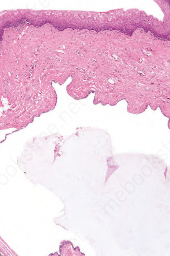
Fig. 12.174 Mucinous cyst: lowpower view of mucincontaining cyst. Note the nonkeratinizing surface epithelium. By courtesy of C. Crum, MD, Brigham and Women’s Hospital and Harvard Medical School, Boston, USA.
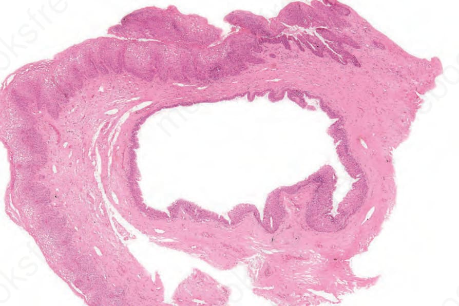
Fig. 12.177 Periurethral cyst: low-power view of unilocular cyst. By courtesy of C. Crum, MD, Brigham and Women’s Hospital and Harvard Medical School, Boston, USA.