Idiopathic calcinosis
Idiopathic calcinosis
Clinical features Genital calcinosis is uncommon. It occurs much more frequently in the scrotum than in the vulva, where it has only seldom been reported.1–7 Very rarely, idiopathic calcinosis may develop in the penis or areola of the nipple.8,9 Lesions present as single or multiple hard nodules in children and young adults (Fig. 12.148). Occasionally, nodules break down and discharge chalky material. Some lesions are polypoid and in this setting the clinical diagnosis is difficult if only a single lesion is present.10–12 Very young children can exceptionally present with single or multiple calcified scrotal lesions secondary to meconium periorchitis.13,14
Pathogenesis and histologic features Although originally thought to represent an idiopathic condition, it more likely that this disorder develops from dystrophic calcification of epidermoid
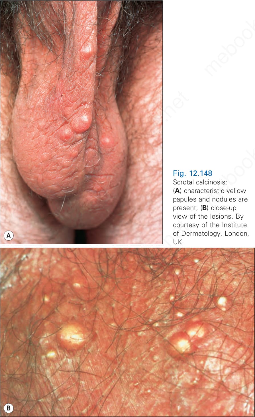
Fig. 12.148 Scrotal calcinosis: (A) characteristic yellow papules and nodules are present; (B) close-up view of the lesions. By courtesy of the Institute of Dermatology, London, UK.