Trichosporosis
Trichosporosis
Clinical features Trichosporosis due to Trichosporon beigelii is a common form of genitocrural and perianal intertrigo in India.1 Predominant symptoms are itching and/ or burning. Scaly papules can accompany the intertrigo. Coexisting dermatophyte, Candida, trichomycosis, and erythrasma infection may be found. Infection rarely occurs elsewhere, including the scalp.2,3
Histologic features The infection involves the hairs but not the surrounding skin. Microscopic examination of hair shafts shows white or brown soft nodules of varying size that can easily be removed.2 The diagnosis is suspected by a KOH preparation to identify variable-sized arthrospores and is confirmed by culture.
497 Infectious diseases
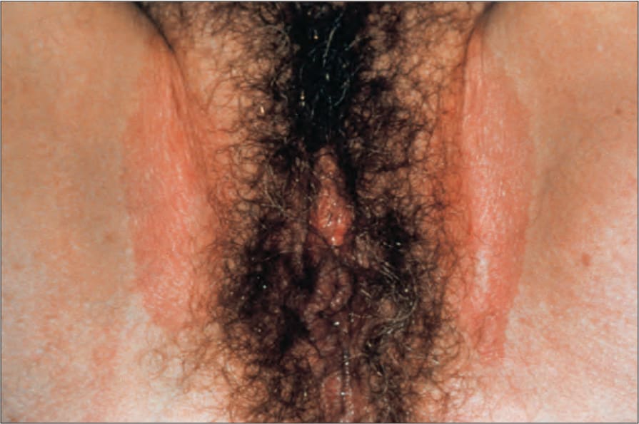
Fig. 12.77 Erythrasma: the flexural distribution and sharply demarcated border are characteristic features. By courtesy of the Institute of Dermatology, London, UK.
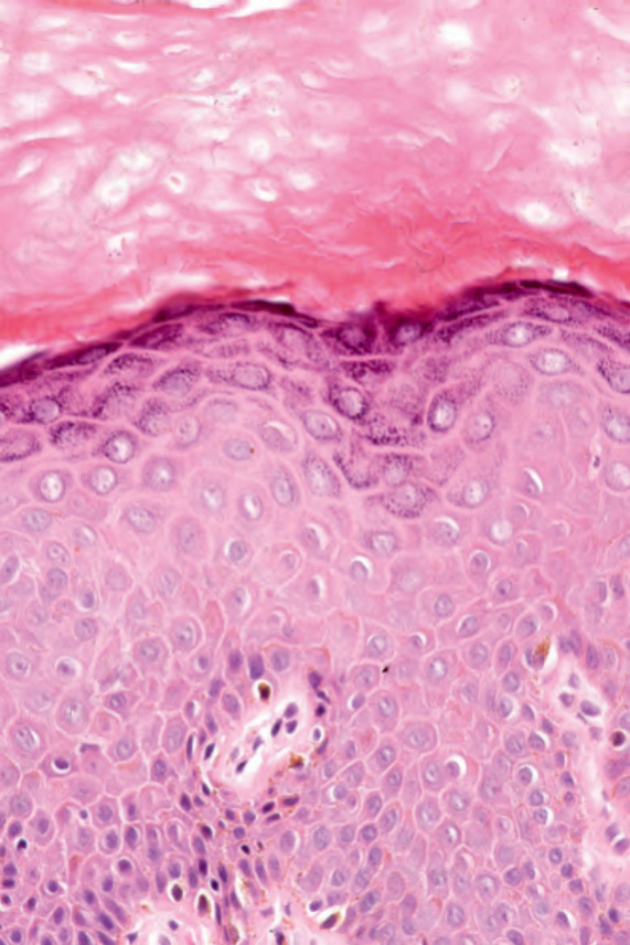
Fig. 12.78 Erythrasma: bacilli are just visible in the upper stratum corneum.
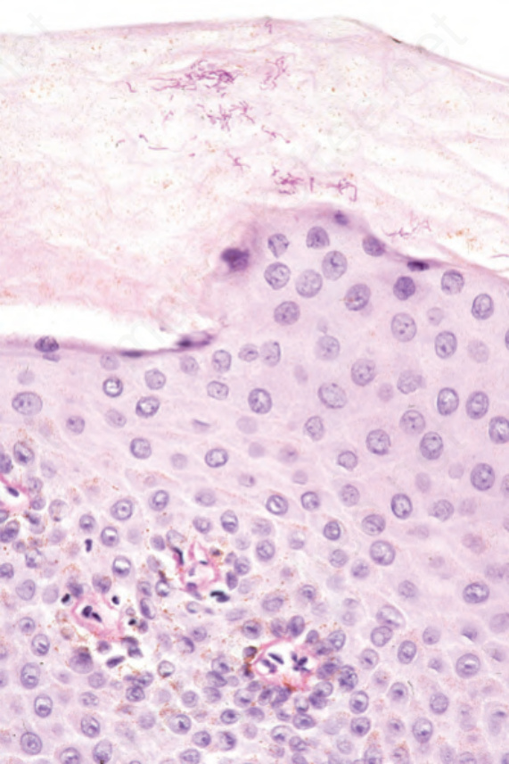
Fig. 12.79 Erythrasma: PAS stain showing elongated bacilli.
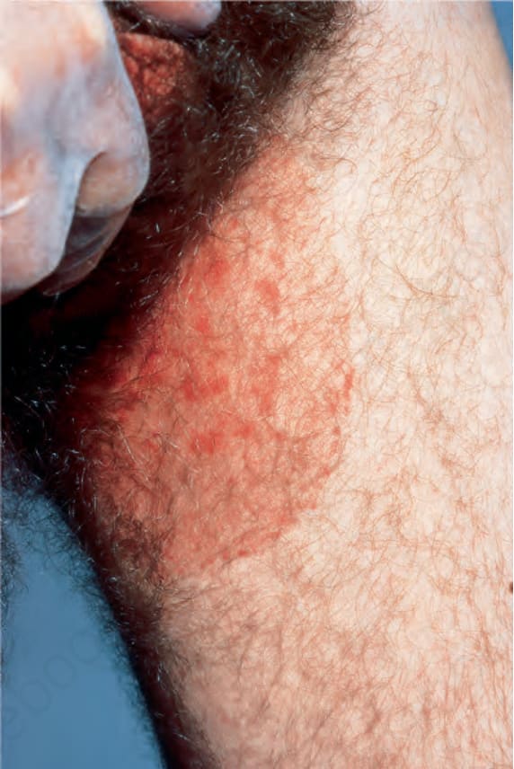
Fig. 12.80 Tinea cruris: there is a large erythematous lesion involving the inner thigh. From Bunker C. Male Genital Skin Disease. Saunders Ltd./Elsevier 2004.
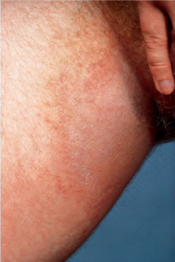
Fig. 12.81 Tinea cruris: note the acute margin. From Bunker C. Male Genital Skin Disease. Saunders Ltd./Elsevier 2004.
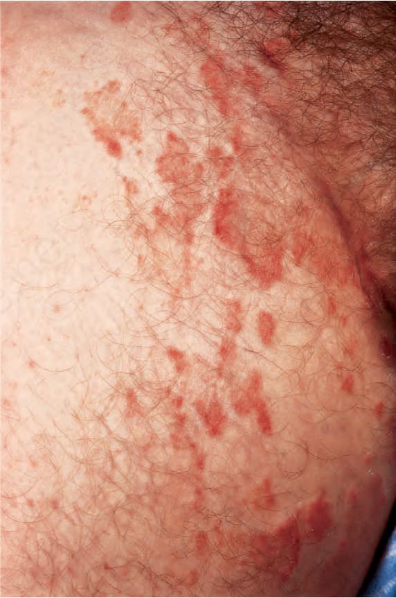
Fig. 12.82 Tinea cruris: close up view. From Bunker C. Male Genital Skin Disease. Saunders Ltd./Elsevier 2004.
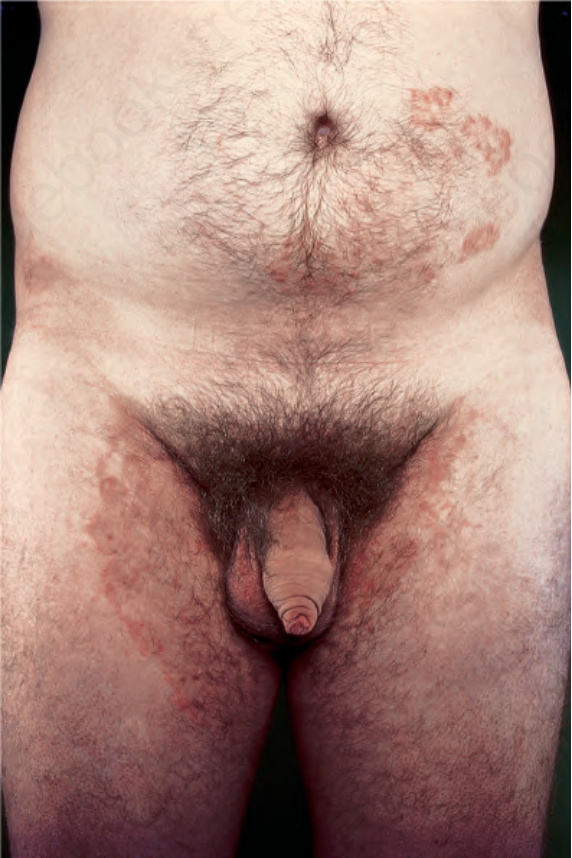
Fig. 12.83 Tinea cruris: in this patient, there is extensive involvement. From Bunker C. Male Genital Skin Disease. Saunders Ltd./Elsevier 2004.
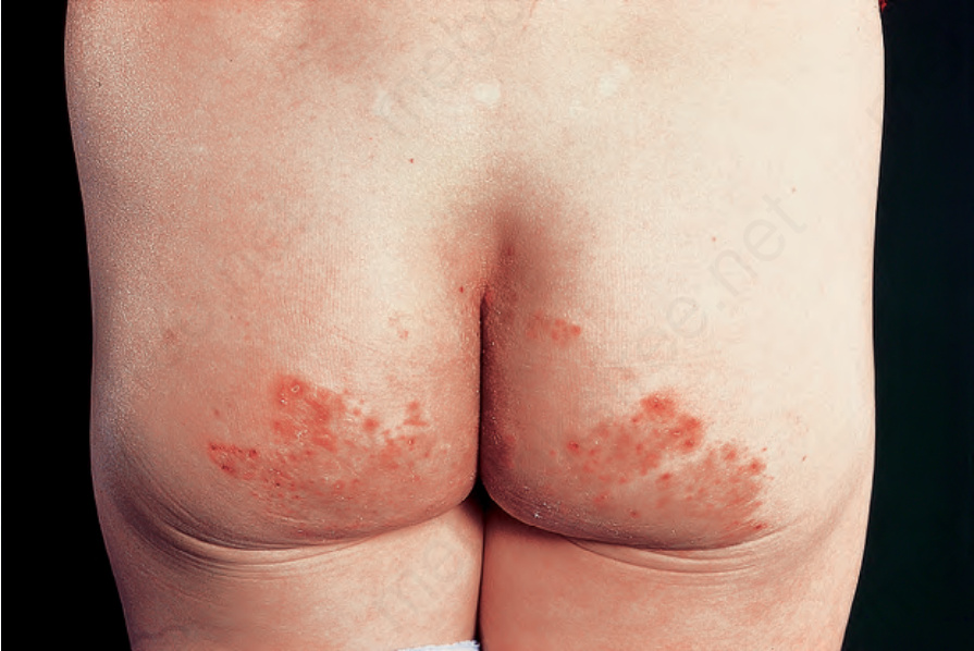
Fig. 12.84 Tinea cruris: note the bilateral involvement of the buttocks.