Myointimoma
Myointimoma
Clinical features This is a rare, recently described tumor involving the corpus spongiosum of the glans penis.1 The original series consisted of adult patients, but a recent small series in children and adolescents has been reported.1,2 Few single case reports have been presented.3–5 Lesions are small (usually less than 1 cm) and asymptomatic. It does not seem to be related to trauma. The behavior is benign with no tendency for local recurrence.1,2
• size larger than 5 cm in diameter,
• infiltrative margins,
• more than 5 mitoses/10 high-power fields (HPF),
• moderate to severe cytological atypia. Tumor necrosis should also be regarded as evidence of malignancy.11
Histologic features Low-power examination reveals a diffuse myointimal proliferation of the blood vessels of the corpus spongiosum of the glans penis in a plexiform growth pattern (Figs 12.297–12.299). The proliferating cells are bland and spindled with abundant pink cytoplasm and vesicular nuclei. A minority of the cells display features more reminiscent of fibroblasts. The background stroma is sclerotic and myxoid. Focal degenerative changes may be seen and mitotic figures are absent.
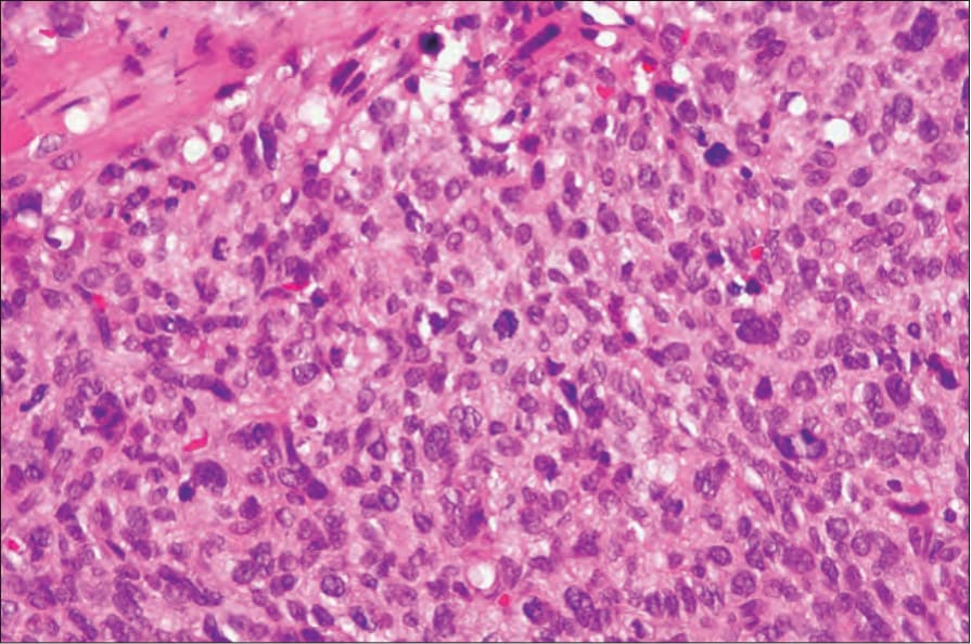
Fig. 12.296 Vulval leiomyosarcoma: high-power view showing a mitotic figure in the center of the field. By courtesy of C. Crum, MD, Brigham and Women’s Hospital and Harvard Medical School, Boston, USA.
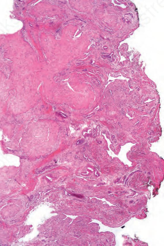
Fig. 12.297 Myointimoma: note the multinodular and plexiform growth pattern.