Viral exanthemata
Viral exanthemata
A variety of viral infections may present with cutaneous eruptions. Although some viruses are associated with an eruption with distinctive clinical features, others are affiliated with a non-specific maculopapular dermatosis (Table 8.1). Exceptionally, exanthemata may represent a primary cutaneous infection. More commonly, the clinical features are a manifestation of an immune response, such as an immune complex disease or a cell-mediated hypersensitivity reaction, to an infection at an extracutaneous site.
Biopsy of a viral exanthem often shows a superficial perivascular lymphocytic infiltrate. Some cases may show epidermal pathology such as interface changes with dyskeratotic cells. The histologic features are entirely non-specific, and distinction from hypersensitivity reactions (e.g., a drug eruption) is impossible without clinical (often including serological investigation) correlation. Viruses that infect cutaneous sites may be visualized by light microscopy including immunohistochemistry, or demonstrated by viral culture, immunological testing, PCR or DNA hybridization. Skin manifestations of viral infections are described in detail in Chapter 18.
Pathogenesis and histologic features The pathogenesis of this condition is unknown.
Histologically, the eruption is characterized by a superficial and deep lymphohistiocytic infiltrate accompanied by conspicuous eosinophils. The epidermis is hyperkeratotic and there is mild acanthosis frequently
Access ExpertConsult.com for the complete list of references
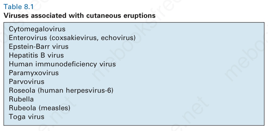
Table 8.1 Viruses associated with cutaneous eruptions