Harlequin ichthyosis
Harlequin ichthyosis
A
Clinical features Harlequin ichthyosis (harlequin fetus, ichthyosis fetalis, ichthyosis congenita gravis) is a distinct severe subtype of autosomal recessive congenital ichthyosis, where babies are born with a fissured ‘armor-plated’ skin (Fig. 3.19).1–4 Characteristically, ectropion and eclabium are very severe. Constricting bands of digits can lead to autoamputation; ears and nose may be malformed.1 The harlequin fetus still has a high mortality due to infections, and respiratory and feeding difficulties accompanied by excessive fluid loss.3 However, treatment with retinoids and improved intensive care have certainly improved the prognosis and quality of life. Surviving neonates develop a severe erythroderma reminiscent of CIE, and there are intermediate cases that show considerable improvement of the skin compared with the initial dramatic presentation.5 Antenatal diagnosis is possible by genetic analysis as well as electron microscopy of fetal skin biopsy (fetoscopy).6,7
B
Pathogenesis and histologic features This very rare form of ichthyosis is due to a dramatic loss of function of the LBs, which results from nonsense mutations in the ABCA12 gene.8 Less severe missense mutations are associated with LI or CIE. Intermediate forms are possible.9,10 The ABCA12 gene product belongs to the ATP-binding cassette (ABC) transporter family that encompass a variety of membrane proteins involved in the energy-dependent transport across membranes. In the epidermis, ABCA12 plays an important role in the LB function. As such it is responsible for the transfer of glucosylceramides, which are essential lipids for epidermal barrier formation. Moreover, it transports proteases such as kallikrein 5, 7, or 14 and secretes them into the intercellular space in the stratum corneum.11 These proteases play an important role in desquamation by degrading corneodesmosomes, thus leading to retention hyperkeratosis.12
Other mutations in ARCI been detected in the CYP4F2 gene encoding a cytochrome P450 polypeptide, CERS3 gene encoding a ceramide synthase, and PNPLA1, a member of the patatin-like phospholipase family.32–35 Moreover, less severe missense mutations in the ABCA12 gene are associated with LI or CIE. Of note, nonfunctional severe nonsense mutations of the same gene are responsible for the far more severe ichthyosis form of harlequin ichthyosis (see below).36,37
Histologically, the epidermis is pale and characterized by massive hyperkeratosis (sometimes with lipid deposits) associated with a normal or absent granular cell layer (Fig. 3.20). The hair follicles are usually affected first, during the second trimester.2,7 Parakeratosis may also sometimes be evident.13 Acanthosis is often marked and papillomatosis is sometimes a feature. A sparse mixed inflammatory cell infiltrate can be present in the superficial dermis.7
Ultrastructurally, the harlequin fetus is associated with deficient or morphologically abnormal LBs (including concentrically lamellated forms) and deficient intercellular lipid lamellae within the stratum corneum.1,2,13 Small vesicles, devoid of internal lamellation, may be present in the granular cell layer (and retained in the stratum corneum), but show no association with the keratinocyte cell membranes as is typical of normal LBs.1,13
Histologic features Histologically, the epidermis in ARCI shows marked orthohyperkeratosis, mostly of the compact type (which may be extreme in the collodion baby) and mild acanthosis with a normal or thickened granular cell layer (Fig. 3.18). The hyperkeratosis is much less marked in erythrodermic than in nonerythrodermic forms. Epidermal papillomatosis associated with a psoriasiform appearance has also been documented. A perivascular lymphocytic infiltrate is occasionally a feature.38 Dilatation and tortuosity of the dermal capillaries is sometimes evident. Follicular hyperkeratosis may rarely be seen. The histologic changes cannot be correlated with the underlying genetic defect.
Ultrastructural studies show a variety of features, including defective development of the cornified cell envelopes and electron-dense debris adjacent to the plasma membranes, lipid vacuoles, increased numbers of small and dysmorphic LBs, or membrane packages.39 Cholesterol clefts are found in TG-1 defects.40 Abnormal LBs and elongated membranes in the
Immunohistochemical evidence suggests that these vesicles represent abnormal LBs characterized by an inability to discharge their lipid contents into the intercellular space. Keratin and filaggrin expression have also been shown to be defective.2 In the harlequin fetus, the keratinocytes may display the hyperproliferative keratins K6 and K16 and show an inability to convert profilaggrin to filaggrin.2
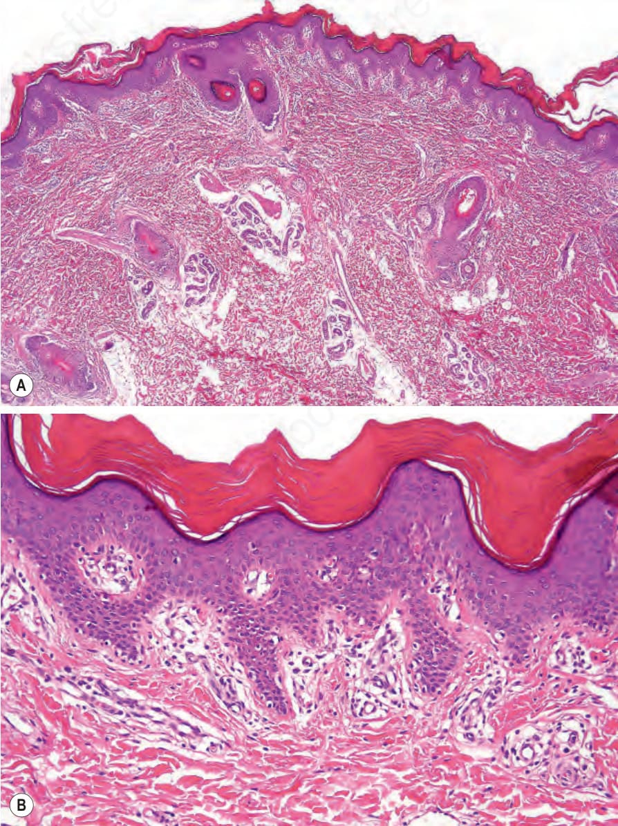
Fig. 3.18 Autosomal recessive congenital ichthyosis: (A) there is very marked orthohyperkeratosis and the epidermis shows papillomatosis; (B) the stratum granulosum is preserved. Note a mild lymphocytic infiltrate.
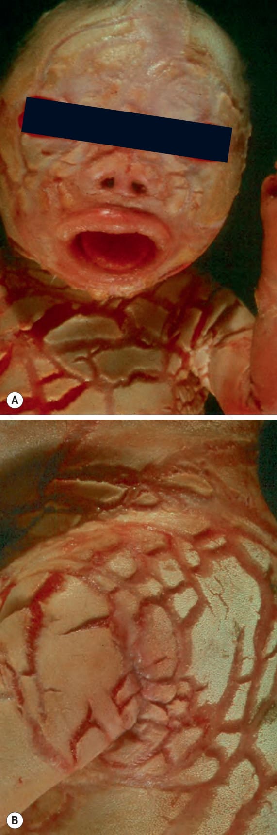
Fig. 3.19 (A, B) Harlequin ichthyosis: the most extreme form of congenital ichthyosis. The scales are very thick and are often referred to as armor-plating. By Courtesey of Sabine Köhler, Stanford University.
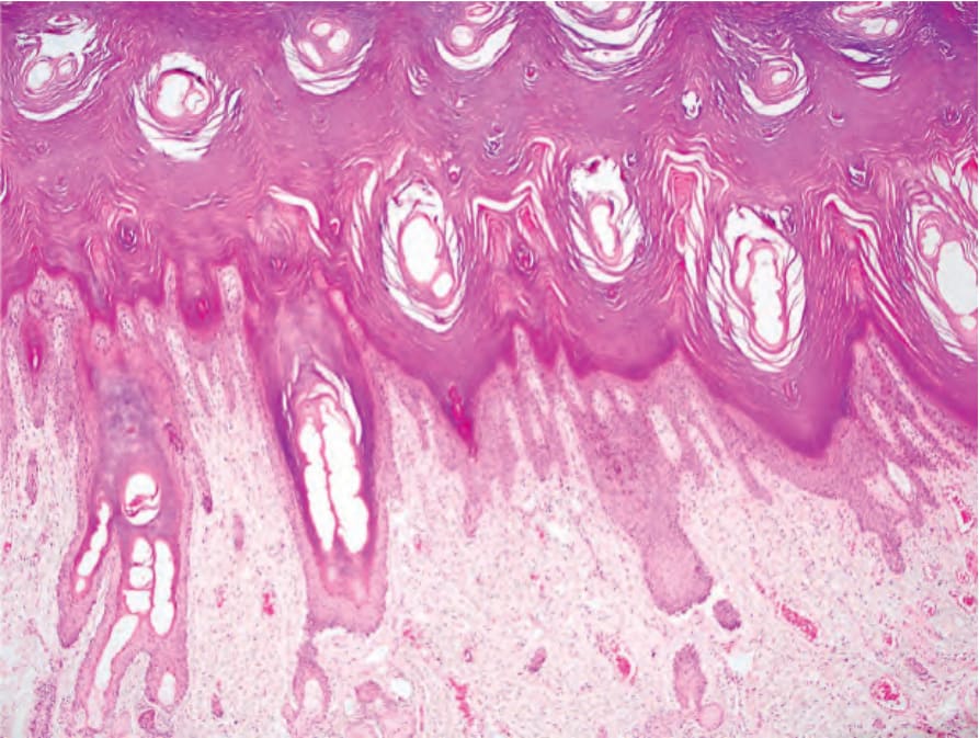
Fig. 3.20 Harlequin ichthyosis: there is massive hyperkeratosis and papillomatous and pale staining epidermis with thinning of the granular cell layer. The dilated spaces in the stratum corneum represent affected hair follicles and sweat ducts. By courtesy of S. Köhler, MD, Stanford University, USA.