Melanocytes
Melanocytes
Melanocytes are pigment-producing cells and are found in the skin, inner ear, choroid and iris of the eye. In skin, melanocytes are located in the
12 The structure and function of skin
A B
basal keratinocyte layer. The ratio of melanocytes to basal cells ranges from approximately 1 : 4 on the cheek to 1 : 10 on the limbs. They appear as vacuolated cells in hematoxylin and eosin stained sections (Fig. 1.35). Ultrastructurally, melanocytes have pale cytoplasm and are devoid of tonofilaments, hemidesmosomes, and desmosomes (Fig. 1.36). They are easily recognized by their specific cytoplasmic organelles (melanosomes) which are derived from the smooth endoplasmic reticulum. Melanosomes are believed to represent a specialized variant of lysosome (Fig. 1.37). The function of melanocytes is the production of melanin, a complex of pigmented proteins that vary in color from yellow to red to brown or black and accounts for the various skin colors within and among races. Melanin protects the mitotically active basal epidermal cells from the injurious effects of ultraviolet light, which accounts for individuals with less pigmentation (fair-haired and light-skinned) having a much greater risk of sunburn and developing
cutaneous malignancies (squamous cell and basal cell carcinomas, and melanoma). The mechanism involves absorbing or scattering ultraviolet radiation and/or its photoproducts. Other functions of melanin include control of vitamin D3 synthesis and local thermoregulation.
In skin and hair, two forms of melanin pigment are produced; eumelanin and pheomelanin. Eumelanin is a brown or black pigment and is synthesized from tyrosine; it is particularly found in dark-colored races, whereas, pheomelanin has a yellow-red color and is synthesized from tyrosine and cysteine; it predominates in Caucasian skin.
Melanocytes also possess melanocyte-specific receptors including melanocortin-1 (MC1R) and melatonin receptors.1 The activation or the inhibition of melanocyte-specific receptors can augment normal melanocyte function, skin color, and photoprotection. Moreover, receptor polymorphisms are known to underlie red hair phenotypes, as well as skin pallor
13 Merkel cells
They may be encountered in normal skin, in lentigines, dysplastic nevi, Spitz nevi, in the café-au-lait macules of neurofibromatosis and in albinism. A key protein involved in melanosome assembly is NCKX5, encoded by the gene SLC24A5.4 Loss of expression of this gene in mice results in marked changes in skin color with loss of pigment. Mature melanosomes of eumelanin are ellipsoidal in shape, while pheomelanin-producing melanosomes are spherical.
or freckles (ephelides).2 Hair graying reflects abnormalities in melanocyte signaling. Notably, Notch transcription factor signaling in melanocytes is essential for the maintenance of proper hair pigmentation, including regeneration of the melanocyte population during hair follicle cycling.3
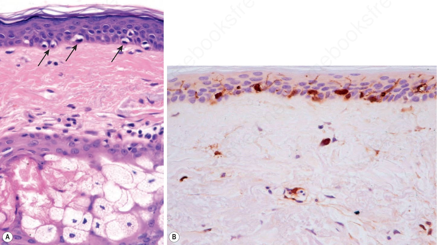
Fig. 1.35 (A, B) Normal epidermis: melanocytes are seen along the basal layer of the epidermis. The cytoplasmic vacuolation is a fixation artifact; (B) melanocytes can be highlighted with S100-protein immunohistochemistry. Note the dendritic processes.
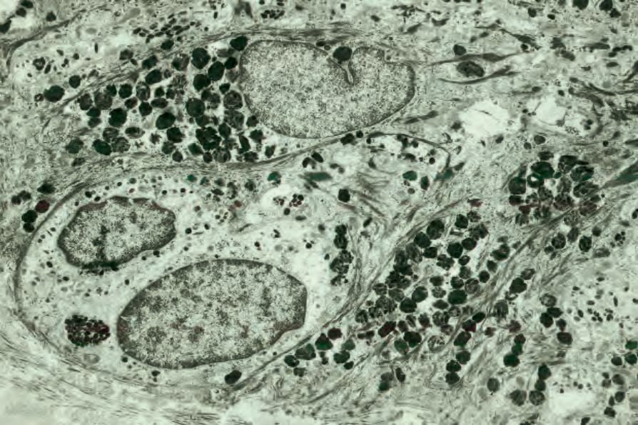
Fig. 1.36 Normal melanocyte: it has abundant pale cytoplasm and scattered solitary melanosomes. Note the absence of tonofibrils and desmosomes.
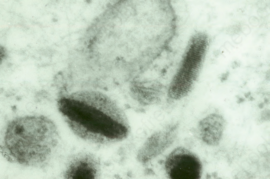
Fig. 1.37 Melanosome: note the typical striated internal structure.
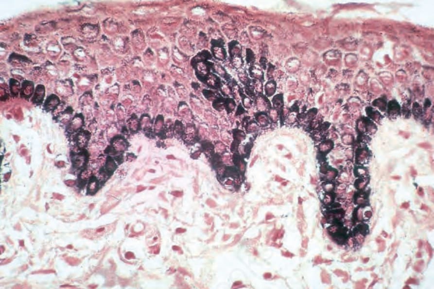
Fig. 1.38 Normal epidermis: this section of black skin has been stained by the Masson– Fontana reaction for melanin. Note the heavy pigmentation, which is present in both melanocytes and keratinocytes.
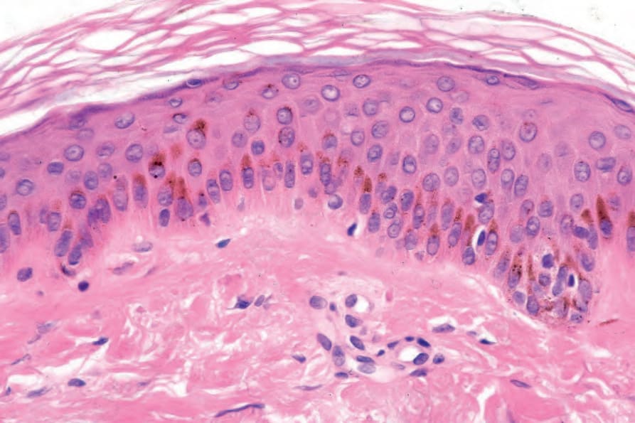
Fig. 1.39 Melanin pigment: actinically damaged skin. Note that the melanin pigment is located in a ‘cap’ overlying the keratinocyte nuclei.
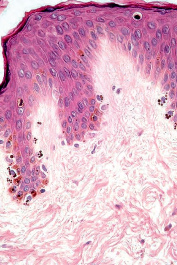
Fig. 1.40 Macromelanosomes: note the large spherical melanosomes in the cytoplasm of the melanocytes.