原理與適應症
- 免疫過氧化酶抗原定位 (Immunoperoxidase antigen mapping) 為 split skin immunofluorescence 的替代法,於石蠟包埋 (paraffin-embedded) 的病灶皮膚切片上,以直接免疫過氧化酶技術判定真皮-表皮分離的層次。
- 定位已知基底膜區成分:keratins 5/14、laminin、type IV collagen 於水疱腔的頂部或底部,藉此判定水疱形成位置為基底內 (intrabasal)、lamina lucida 內或 lamina densa 深層。
各疾病的定位模式 (操作判讀)
- Epidermolysis bullosa simplex:上述所有免疫反應物皆位於水疱腔底部 (floor)。
- Bullous pemphigoid:keratin 位於頂部 (roof),laminin 與 type IV collagen 位於底部 (floor)。
- Dystrophic epidermolysis bullosa、epidermolysis bullosa acquisita、bullous systemic lupus erythematosus:三種免疫反應物皆位於水疱頂部 (roof)。
注意事項與限制
- 許多遺傳性與後天性水疱病中,針對標靶抗原的抗體於石蠟包埋材料上反應不佳,偽陽性與偽陰性常見,使本法不適合常規診斷。
- 遺傳性表皮下水疱病的抗原定位專以冷凍切片 (frozen sections) 進行,結果優異。
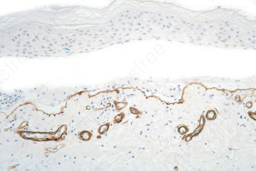
圖 4-7:石蠟包埋免疫過氧化酶抗原定位;於 bullous pemphigoid,type IV collagen 位於水疱底部。
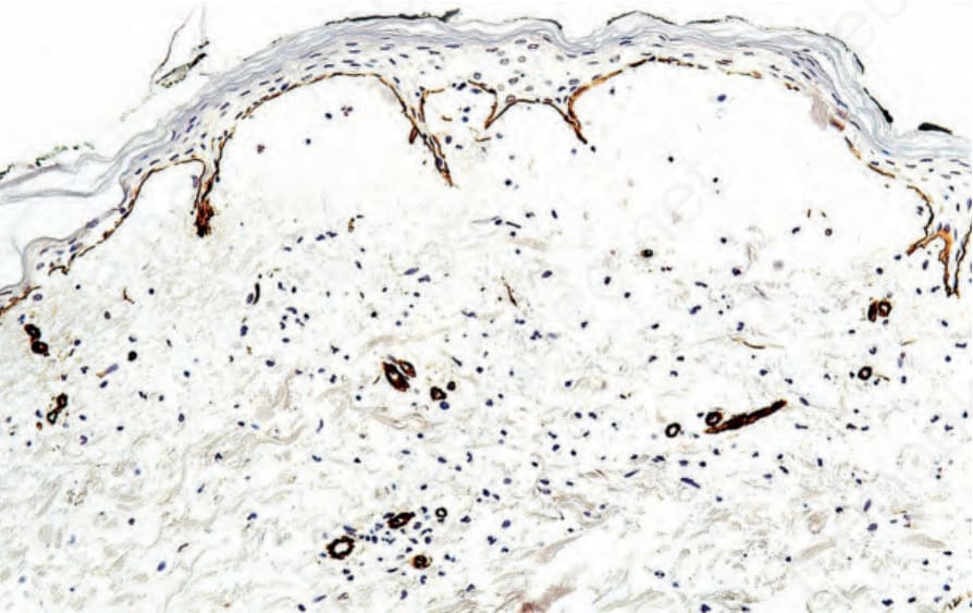
圖 4-8:石蠟包埋免疫過氧化酶抗原定位;於 epidermolysis bullosa acquisita,type IV collagen 位於水疱腔頂部。