染色體核型分析 (Chromosomal karyotyping)
- 可作為初步篩檢,呈現某些腫瘤相關的粗略染色體異常 (gross chromosomal aberrations)。
- 多數皮膚腫瘤體積小,通常不會保留組織專供核型分析。
- 為偵測腫瘤組織染色體異常的歷史性黃金標準 (gold standard)(圖 2.22)。
FISH 原理
- FISH 利用螢光標記探針,與特定基因組 DNA 區域互補並特異性雜交,使該區域可被觀察。
- 目標 DNA 可為中期染色體展開 (metaphase spreads)、間期細胞核 (interphase nuclei)(圖 2.23),或 FFPE 石蠟切片中的細胞核(圖 2.24);探針與目標經變性後接觸數小時至數天,於適當條件下退火 (anneal)。
- 探針鎖定富含重複序列的區域(如著絲點 centromeres)時最易雜交,訊號大而易測;但這些區域無功能性基因,只能辨識拷貝數增加,無法得知特定癌基因/基因座的拷貝數。
- 偵測僅涉及染色體片段的異常(人類癌症常見,含 melanoma)需鎖定獨特(非重複)序列的探針,訊號較小、較難偵測;使用 100–300 kb 較大探針即可在石蠟切片偵測獨特序列。
- 優點:即使存在大量正常細胞,只要能形態學辨識腫瘤細胞即可偵測異常。
- 限制:組織切片中偵測異型合子缺失 (heterozygous deletions) 較難(核截斷 truncation 造成訊號隨機遺失);倍體性 (ploidy) 增加可模擬目標基因座增益。
- 對策:同時雜交多個不同顏色探針(空間補償)+ 分析更多細胞(統計補償);以同染色體上另一顏色的著絲點探針作對照可控制非整倍體 (aneuploidy)。
- 經典範例:比較 17q 上 HER2 基因座訊號與 centromere 17,可區分第 17 號染色體拷貝數增加 vs. HER2 基因座的特異性擴增 (amplification)。
斷裂分離探針 (Break-apart probe) 技術
- 以側翼於易位斷點 (breakpoint) 兩側的探針進行 FISH,可呈現該基因座重排(圖 2.25);對具反覆性易位的造血惡性腫瘤(如 large cell anaplastic lymphoma)及某些可侵犯皮膚的軟組織腫瘤具診斷價值。
- 側翼探針以兩色(如綠、紅)標記:完整染色體上兩訊號並列,光譜重疊產生黃色訊號(每條正常染色體一個)。
- 以 clear cell sarcoma(22q12 的 EWSR1 重排)為例:完整 22 號染色體呈黃色訊號;重排後著絲點探針保留於衍生 22 號,端粒探針 (telomeric probe) 轉移至接受染色體(clear cell sarcoma 為第 12 號),形成分開的紅、綠訊號。
- 關鍵限制:僅指出基因座發生重排,無法指出染色體夥伴與基因身分;不同腫瘤的不同易位可涉及同一基因座,須謹慎判讀。
- 例:EWSR1 在 clear cell sarcoma、Ewing sarcoma、extraskeletal myxoid chondrosarcoma、angiomatoid fibrous histiocytoma 及部分 myxoid liposarcoma 皆可重排(圖 2.26);須與臨床及組織學特徵對照。
其他原位雜交 (In situ hybridization) 變化
- FISH 缺點:一次只能看少數基因座、需計數大量細胞核而耗時(已由電腦化計數演算法部分克服)。
- CISH(顯色性探針):以光學顯微鏡即可觀察訊號,適合偵測基因座擴增;技術上可用斷裂分離策略偵測易位,但實務判讀困難。
- CISH 最常見用途為直接偵測感染相關核酸,如 HPV 或 Epstein-Barr 病毒;HPV 可分型為高風險型(16、18)或低風險型(6、11)。改良版亦可偵測組織中的 messenger RNA。
- 原位雜交(螢光或顯色)最適合呈現:特定基因的擴增、特定基因的重排、「外來」(感染相關)DNA/RNA 的存在。
- 改良技術 multiplex FISH、光譜核型分析 (spectral karyotyping)、全染色體塗染 (whole chromosomal painting) 可為每條染色體個別著色,便於核型分析及辨識融合/衍生染色體,對複雜核型判讀極有幫助;電腦輔助偽彩色 (pseudocoloration) 可促進半自動化核型分析。

圖 2-25:斷裂分離 FISH 技術 (break-apart FISH technique):圖示與 clear cell sarcoma 相關的 12;22 易位。(A) EWSR1 完整時,探針雜交至基因兩側的著絲點側(紅)與端粒側(綠),並列產生黃色訊號,故未重排細胞可見兩個黃色訊號(代表兩條未重排的第 22 號染色體);(B) 發生重排時(與第 12 號染色體的平衡易位),著絲點探針(紅)保留於衍生第 22 號,綠色探針轉移至衍生第 12 號,故核中一個黃色訊號代表完整 22 號,衍生 12 與 22 號分別以單一綠、紅訊號自由分離。
Fig. 2.25 Break-apart FISH technique: the 12;22 translocation associated with clear cell sarcoma is depicted. (A) When the EWSR1 locus is intact, the probes hybridize to the centromeric (red) and telomeric (green) regions flanking the gene. The spectral overlap of the two signals in juxtaposition produce a yellow signal. Thus in cell lacking rearrangement of this locus, two yellow signals are present, representing the two copies of chromosome 22 lacking rearrangement (right); (B) When rearrangement occurs, such as the balanced translocation with chromosome 12 depicted here, the centromeric probe (red) is retained by the derivative chromosome 22 while the green probe is transferred to the derivative chromosome 12. Thus in the nuclei one yellow signal indicates the intact chromosome 22 while the derivative 12 and 22 chromosomes segregate freely as single green and red signals, respectively (right).
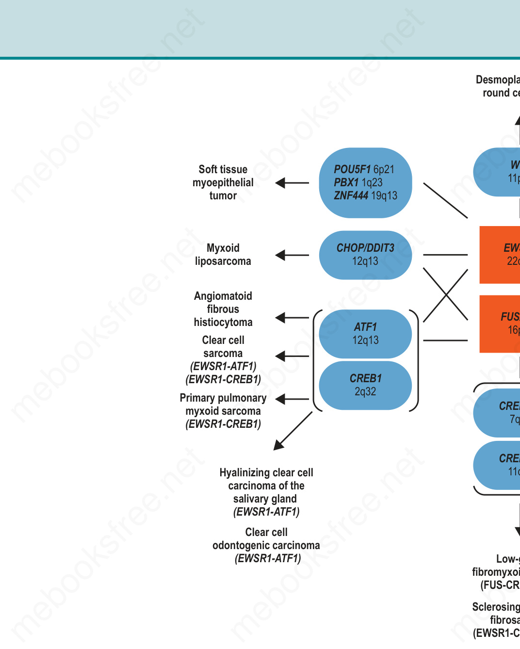
圖 2-26:涉及 EWSR1 及其同源基因 FUS 的多重易位:EWSR1 與 FUS 常可互相取代,且皆參與和多個基因的平衡易位,形成各式腫瘤。由於 FISH 僅指出單一基因座(如 EWSR1)發生重排,而不提供融合夥伴 (fusion partner) 的資訊,結果須在臨床與形態學背景下謹慎判讀;有時須以 RT-PCR 等技術確認融合夥伴。
Fig. 2.26 Multiple translocations involve EWSR1 and the homologous gene, FUS: both EWSR1 and FUS can often substitute for one another and both are involved in balanced translocations with multiple genes resulting in a variety of neoplasms. Since FISH only indicates that a single locus, such as EWSR1, is rearranged and nothing about the fusion partner, results must be interpreted carefully within the clinical and morphologic context of a tested case. Sometimes techniques such as RT-PCR must be used to verify the fusion partner.