Botryomycosis
Botryomycosis
Bacterial induction of disease is minimized by autoclaving or irradiating feed, treating the water (acidification or chlorination), and maintaining a high barrier. However, not all investigators follow protocols, so accidental infection of mice with human pathogens can occur. Some bacterial infections can become quite serious in the skin of mice, for example, Staphylococcus spp. induced botryomycosis. Here, mice can present with massive abscesses often around the head from which pure cultures of the bacteria can be isolated (Fig. 36.7). These infections can spread to the lungs with a miliary distribution giving the appearance of metastatic cancer. While the lesions may present as a skin lesion, entry into the body usually can be found coming through ulcerations in the epithelium surrounding the molars, which are often induced by impaction with food material and hairs (from grooming). Osteomyelitis in the maxilla spreads, resulting in prominent swelling of the face which may rupture at the skin surface. This lesion can be a major part of the phenotype of some targeted mutant mice, notably those with deficiencies in cytochrome b-245, beta polypeptide (Cybbtm1Din)2 or plasminogen activator, urokinase (Plautm1Mlg),3 which both affect the immune system. Sentinel mice with aglobulinemia were reported to develop these lesions.4 Staphylococcal infections have resulted in botryomycosis in cut wounds in pathogen-free colonies.5,6
most common types of models, can be simple single gene Mendelian traits (such as coat color) or complex polygenic traits. Those with similar phenotypes due to mutations in different genes are often caused by genes that function in the same molecular pathway. As such, rather than present an encyclopedic listing of models, representative ones that fall into known or novel and evolving pathways that illustrate the power of mouse models to unravel the complexities of skin disease in mammals, which includes humans, are presented.
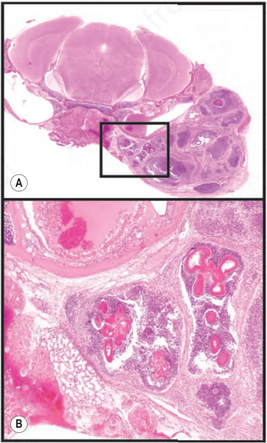
Fig. 36.7 This is a 91-day-old female mouse with botryomycosis. (A) Note the pink immune complexes (Splendore-Hoeppli phenomenon) surrounded by neutrophils and a wall of macrophages and fibroblasts. (B) Lower panel is enlargement of boxed area.