Cherry angioma
Cherry angioma
Clinical features Cherry angiomas (senile angiomas, Campbell de Morgan spots), regarded as variants of capillary hemangiomas, are very common and present as multiple tiny red papules on the trunk and upper limbs of the middle aged and
1832 Connective tissue tumors
elderly.1 An unusual case of cherry angiomas with segmental dyschromatosis and blue nevi has been reported.2
Pathogenesis and histologic features The pathogenesis is unknown. HRAS and KRAS mutations have been found in a small number of cases indicating involvement of the ERK pathway.3 A case of eruptive lesions associated with topical nitrogen mustard therapy, another associated with exposure to bromides, a familial nevus flammeus with early-onset cherry angiomas and a case associated with familial cerebral cavernous malformations have been documented.4–9
Histology shows a small polypoid lesion with an epidermal collarette and multiple lobules of dilated and congested capillaries in the papillary dermis (Fig. 35.469). In a single case, lesions of cherry angioma were colonized by intravascular large B-cell lymphoma.10
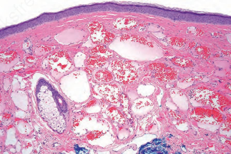
Fig. 35.469 Cherry angioma: there are widely dilated, congested vessels in the superficial dermis.
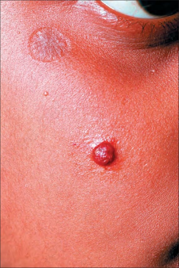
Fig. 35.470 Lobular capillary hemangioma: a typical raised red nodule on the face of a young female patient. By courtesy of M.M. Black, MD, St Thomas’ Hospital, London, UK.