Nevi of the breast
Nevi of the breast
Nevi can occur anywhere on the breast, including in and around the nipple. Unusual histologic features appear to be more common in young adults than in elderly patients.1
Histologic features An analysis of 101 nevi of the breast demonstrated that in comparison to 97 nevi at conventional sites, nevi of the breast more frequently display limited upward migration of melanocytes above the basal layer, presence of random melanocytic cytological atypia, and dermal fibroplasia.1,2 Histologic features of nevi of breast are similar in both genders (Figs 25.75–25.78)2 Nests of melanocytes along the dermal–epidermal junction are variably sized.1 Confluence of nests which are sometimes dyscohesive can be seen. Melanocytes are enlarged with clear to dusty cytoplasm, and dendritic forms may sometimes be seen. Random cytological atypia is frequent and can also be observed in the melanocytes of the papillary dermis.1 The deeper dermal component shows maturation and is generally unremarkable with lack of mitosis activity.
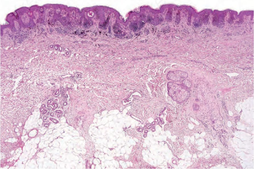
Fig. 25.75 Nevus of breast: there is architectural disorder, pigment incontinence, and a superficial perivascular lymphocytic infiltrate.
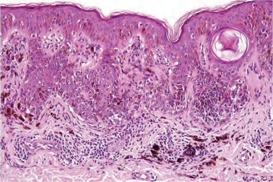
Fig. 25.76 Nevus of breast: note the heavily pigmented lentiginous and nested junctional component.