Fungal infections
Fungal infections
Oral candidiasis is often a sign of progression of HIV infection. Angular cheilitis and onychomycosis are frequent manifestations of candidiasis in patients with severe HIV-induced immunosuppression.1
AIDS defining malignancies (ADMs) include invasive cervical carcinoma, Kaposi sarcoma (KS), and non-Hodgkin lymphomas (NHLs). The incidence of these has declined in well-resourced settings with accessibility to effective ART, although the incidence of non-ADMs did not.6 However, in Africa, KS is an increasing public health problem.7
The most common presentation of KS is multifocal macules or nodules. Extracutaneous KS occurs in 50% and can occur in the absence of skin disease.8 KS is associated with infection with human herpesvirus-8 (HHV-8). Other HHV-8 associated neoplasms are primary effusion lymphoma, a B cell lymphoma and plasmablastic lymphoma. Multicentric Castleman disease (MCD) is associated with HHV-8, and untreated patients are at high risk of developing large B-cell lymphoma (LBCL).9
988.e1 Neoplasia
988.e2 Human immunodeficiency virus (HIV) and acquired immunodeficiency syndrome (AIDS)-associated cutaneous diseases
988.e3 Neoplasia
AIDS-associated NHLs are predominantly of B-cell type. The spectrum includes diffuse LBCL, Burkitt lymphoma, and plasmablastic lymphoma.10 Primary skin involvement is rare, but secondary cutaneous dissemination from a nodal or visceral NHL occurs.11,12
Surprisingly, mycosis fungoides (MF) may rarely develop in patients with HIV and should be distinguished from the uncommon benign epidermotropic CD8+ T-cell infiltrate that has been reported in HIV.13 The latter condition may mimic MF clinically and pathologically.14
989 Immune reconstitution inflammatory syndrome
IRIS-associated infections IRIS-associated noninfectious
Bacillary angiomatosis Blastomycosis Cryptococcus Cytomegalovirus Crusted scabies Dermatophytosis Epidermodysplasia verruciformis Fusariosis Herpes simplex Herpes zoster Histoplasmosis Kaposi sarcoma Leishmaniasis Leprosy Molluscum contagiosum Mycobacterium avium complex Paracoccidioidomycosis Pityriasis versicolor Secondary syphilis Sporotrichosis Strongyloidiasis – pruritus, rash Talaromycosis Tuberculosis – scrofuloderma Warts
Alopecia universalis Acne Eosinophilic folliculitis Erythema elevatum diutinum Lupus erythematosus –
systemic and discoid Mid-dermal elastolysis Non-Hodgkin lymphoma Papular pruritic eruption Psoriasis Pyoderma gangrenosum Reactive arthritis Sarcoidosis Seborrheic dermatitis Sweet syndrome Urticaria
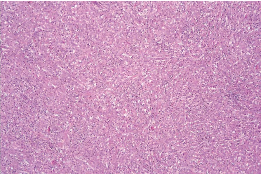
Fig. 19.33 HIV-associated Mycobacterium avium intracellulare infection: within the dermis is a diffuse macrophage infiltrate.
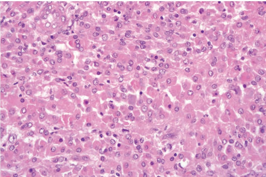
Fig. 19.34 HIV-associated Mycobacterium avium intracellulare infection: the macrophages have intensely eosinophilic cytoplasm and uniform round vesicular nuclei.
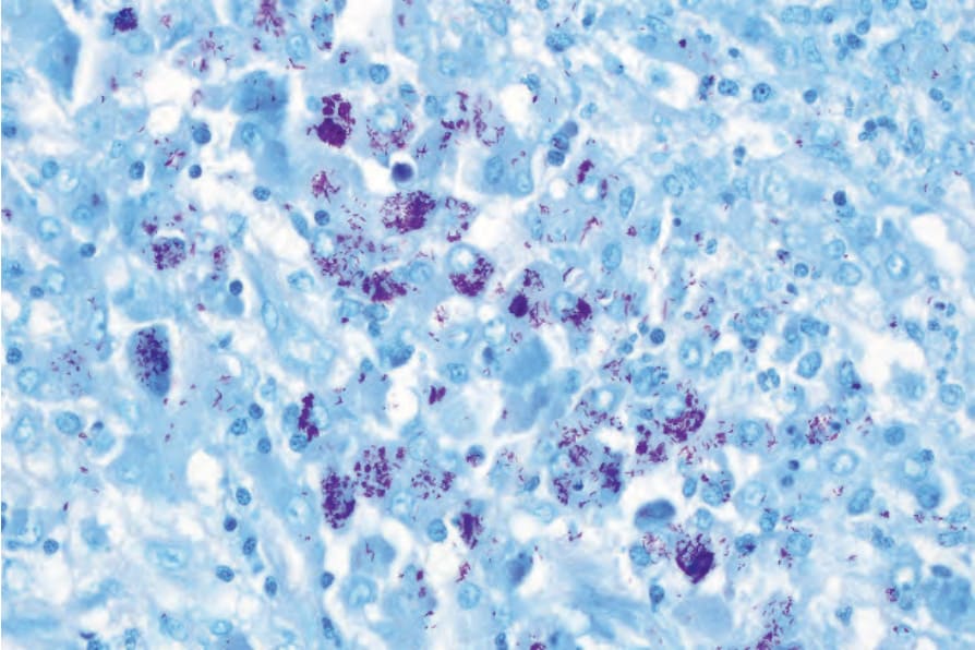
Fig. 19.35 HIV-associated Mycobacterium avium intracellulare infection: an acid-fast stain reveals numerous bacteria.
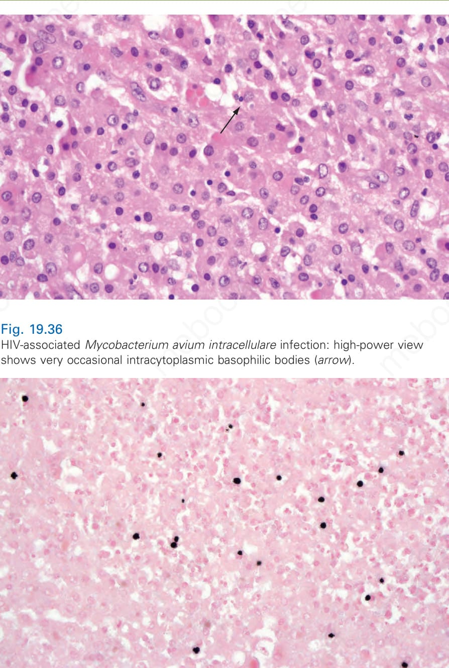
Fig. 19.36 HIV-associated Mycobacterium avium intracellulare infection: high-power view shows very occasional intracytoplasmic basophilic bodies (arrow).
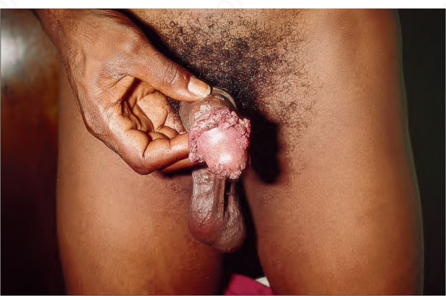
Fig. 19.37 HIV-associated Mycobacterium avium intracellulare infection: a von Kossa stain is positive, confirming the presence of malakoplakia. In HIV-positive patients, multiple pathology is often present in a biopsy specimen.

Fig. 19.38 Genital condylomata and HIV infection: genital and perianal warts may sometimes be associated with exuberant growth in this condition. By courtesy of C. Furlonge, MD, Port of Spain, Trinidad.
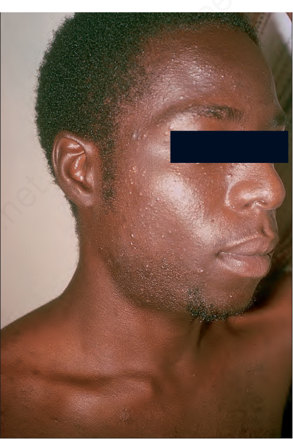
Fig. 19.39 Molluscum contagiosum and HIV infection: this is commonly seen and particularly affects the face. Lesions are typically numerous. By courtesy of C. Furlonge, MD, Port of Spain, Trinidad.
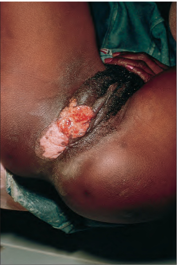
Fig. 19.40 Herpes simplex and HIV infection: there is gross destruction of the vulva with involvement of the thigh. By courtesy of C. Furlonge, MD, Port of Spain, Trinidad.
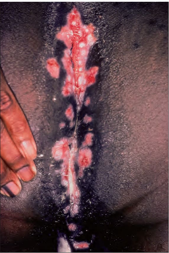
Fig. 19.41 Herpes simplex and HIV infection: severe perianal disease has spread to involve much of the perineum. By courtesy of C. Furlonge, MD, Port of Spain, Trinidad.
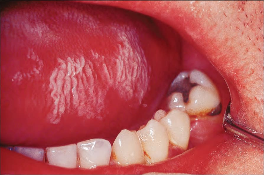
Fig. 19.42 Oral hairy leukoplakia: characteristic delicate linear lesions on the tongue. By courtesy of P.R. Morgan, MD, Institute of Dermatology, London, UK.
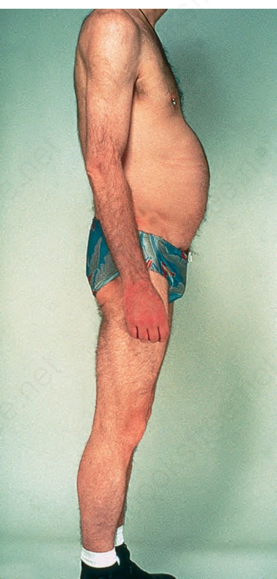
Fig. 19.43 Lipodystrophy associated with HIV infection: there is loss of the fat of the extremities. Note the deposition of fat around the abdomen. By courtesy of D. McGibbon, M.D, Institute of Dermatology, London, UK.
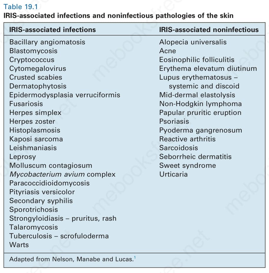
Table 19.1 IRIS-associated infections and noninfectious pathologies of the skin