Favus
Favus
giant cells) and marked fibrosis, which sometimes resembles folliculitis keloidalis.
Clinical features This uncommon pattern of tinea capitis is seen in the Middle East, South Africa, and Greenland, and sporadically elsewhere. It is characterized by cup-shaped crusts, or scutula, around the ostia of hair follicles.1–5 The hair penetrates the crust and is not necessarily broken off or shortened. The crusts may become confluent. Permanent and scarring alopecia occurs (Fig. 18.269). Removal of the scutula leaves an erythematous oozing base. The hairs show a gray-green fluorescence under Wood lamp.
Histologic features Favus is caused by Trichophyton schoenleinii, which invades the hair and produces air spaces, but arthrospores are not seen.1 Relatively little damage occurs to the hair shafts. Fungal hyphae and spores are seen in the scutula, which rests on the acanthotic stratum spinosum around the follicular ostia. The underlying dermis shows a mixed inflammatory infiltrate (including
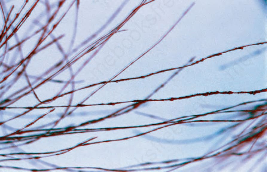
Fig. 18.269 Favus: scarring alopecia due to infection by Trichophyton schoenleinii. By courtesy of R.A. Marsden, MD, St George’s Hospital, London, UK.
Fig. 18.270 Black piedra: there are numerous tiny black nodules attached to hair shafts. By courtesy of the late C. Kalter, MD, Walter Reed Medical Center, Washington, USA.