Strongyloidiasis
Strongyloidiasis
Clinical features Strongyloidiasis refers to infection with the nematode, Stongyloides stercoralis.1,2 An estimated 30 to 100 million people worldwide are infected.3 Although the parasite is present throughout tropical and temperate regions, a high proportion of the population is infected in some tropical countries. S. stercoralis has three life cycles. In the direct development cycle, rhabditiform
Histologic features Skin biopsy reveals intradermal filariform larvae, which may be seen in both longitudinal section and in cross section (Fig. 18.400).3,11 These larvae measure 300–600 µm in length and 10–20 µm in diameter. Minute double
lateral alae may be visible on transverse sectioning.1 Invasion through the dermal vessel walls is associated with vasculitic alterations, with perivascular red cell and fibrin extravasation.7,11
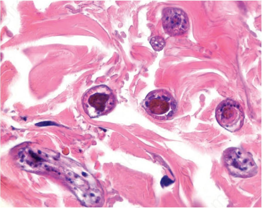
Fig. 18.400 Strongyloidiasis: rhabditiform larvae are present in the dermis of this patient with disseminated infection in the context of underlying AIDS.