Mesothelial cyst
Mesothelial cyst
Clinical features Mesothelial cyst (cyst of the canal of Nuck) presents as a lesion varying in size from less than 1 cm to 5 cm or more. It arises on the upper and lateral aspect of the labium majus at the level of the insertion of the round ligament. Some cases are associated with an inguinal hernia. Simple excision is curative.
Histologic features Microscopic examination reveals a unilocular cavity lined by a single layer of flattened mesothelial cells.1
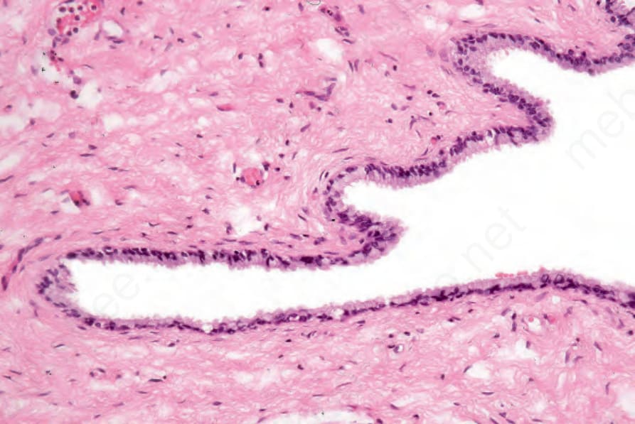
Fig. 12.175 Mucinous cyst: the cyst is lined by mucin-secreting epithelium. By courtesy of C. Crum, MD, Brigham and Women’s Hospital and Harvard Medical School, Boston, USA.