Sclerosing lipogranuloma
Sclerosing lipogranuloma
Clinical features Sclerosing lipogranuloma (paraffinoma) may involve the scrotum, penis, and exceptionally the epididymus.1–3 It usually represents a tissue response
515 Miscellaneous conditions
to exogenous material (usually paraffin, silicone and even mineral oil and Vaseline) and is seen most commonly in the paratesticular area secondary to the injection of size-enhancing materials.4,5 A very small number of cases of sclerosing lipogranuloma appear to be primary, are more common in Japan, and have predilection for the genitourinary system but can occur elsewhere including the rectum.6–8 Granulomatous penile nodules (Tanko nodules) have been described due to the insertion of glass spheres under the penile skin.9
Histologic features There is a foreign body-type granulomatous reaction in the dermis. ‘Lipid’ vacuoles are surrounded by dense fibrous tissue. Rare extensive necrosis in a case of primary sclerosing lipogranuloma has been documented.8
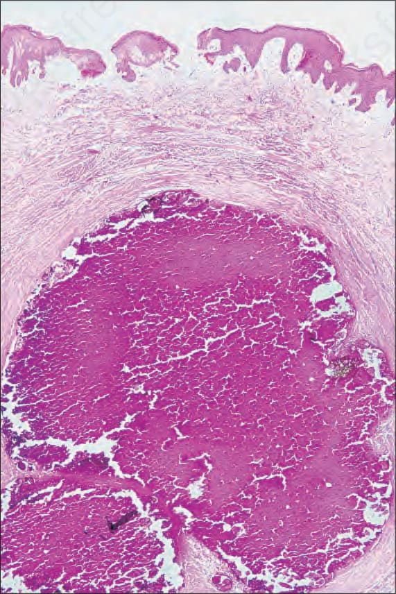
Fig. 12.149 Scrotal calcinosis: the calcium deposits stain purple with hematoxylin and eosin. Sometimes a preexistent epidermoid cyst can be identified.
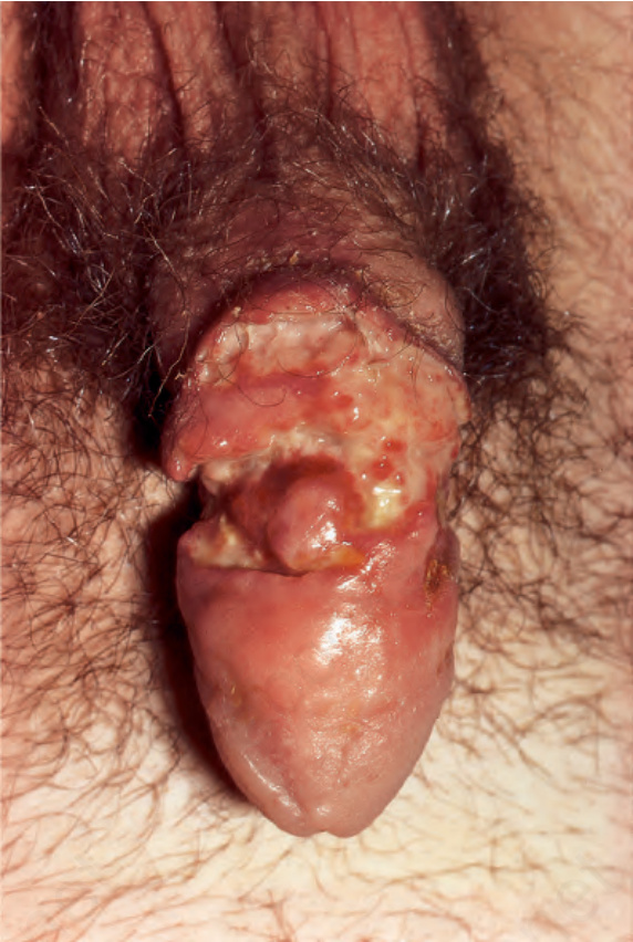
Fig. 12.150 Dermatitis artifacta: there is gross destruction of the penile shaft. From Bunker C. Male Genital Skin Disease. Saunders Ltd./Elsevier 2004.
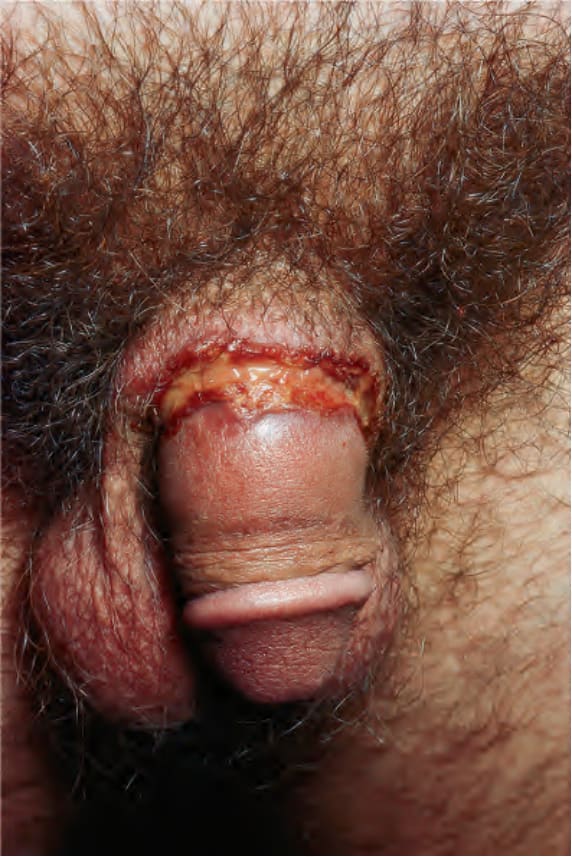
Fig. 12.151 Dermatitis artifacta: note the circumferential ulceration. Reproduced with permission from Bunker C.B. and Mallon E., Management of penile erosions and ulcers, Postgraduate Doctor Middle East, 1998;21:163–168. Copyright © Professional Managerial and Healthcare Publications Limited. From Bunker C. Male Genital Skin Disease. Saunders Ltd./Elsevier 2004.
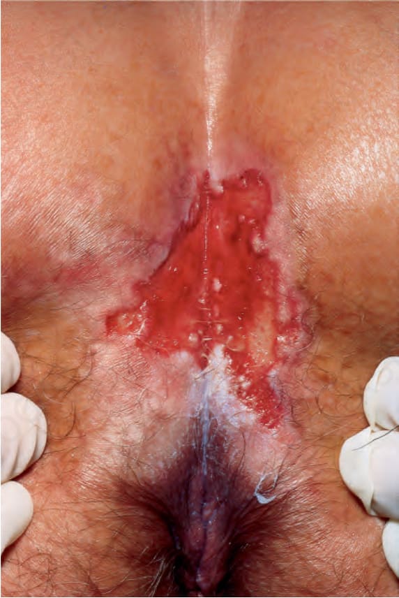
Fig. 12.152 Dermatitis artifacta: this is a view of the natal cleft. Note the central ulceration and surrounding lichenification. From Bunker C. Male Genital Skin Disease. Saunders Ltd./Elsevier 2004.