Pubic hair
Pubic hair
Pubic hair appears during puberty as vellus hair that is focally replaced by terminal hair. Men have a different pattern of pubic hair than women, but in practice it is one of degree. The distribution of hair and pubic hair varies widely between men.1 Generally, the abdominal wall, pubic mound, groins, scrotum and perineum are hairy, but the natal cleft, perianal skin, distal penile shaft, prepuce, and glans are hairless.
In females, pubic hair development starts at puberty on the mons pubis and labia majora. The adult distribution with triangular pattern on the mons, with extension to the labia majora and on to the thighs is usually complete by the age of 16–17 years.
Genital tubercle
Urogenital fold Labioscrotal swelling
Urogenital membrane
Cloacal fold Cloacal membrane
Perineum
Anal membrane
Anal fold
475 Anogenital mammary-like glands
Urogenital membrane breaks down
6th week Early 7th week 7th week A
6th to 7th week 14th week
Endoderm
Urethral groove Urethral plate Urethral fold Penile urethra
3rd month
Epithelial invagination
B
A
B
476 Diseases of the anogenital skin
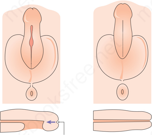
Fig. 12.11 Formation of the external genitalia in males and females. (A) The external genitalia form a pair of labioscrotal folds, a pair of urogenital folds and an anterior genital tubercle. Male and female genitalia are morphologically indistinguishable at this stage. (B) In males, the urogenital folds fuse and the genital tubercle elongates to form the shaft and glans of the penis. Fusion of the urogenital folds encloses the definite urogenital sinus to form most of the penile urethra. A small region of the distal urethra is formed by the invagination of ectoderms covering the glans. The labioscrotal folds give rise to the scrotum. From Bunker C. Male Genital Skin Disease. Saunders Ltd./Elsevier 2004.
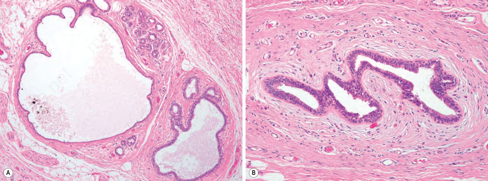
Fig. 12.12 Anogenital mammary-like glands. (A) Medium-power view showing glands with focal cystic dilatation; (B) high-power view showing a double layered cell wall. Focally, there is a suggestion of apocrine differentiation. By courtesy of D. Kazakov, Charles University Medical Faculty Hospital, Pilsen, Czech Republic.