Keloid
Keloid
It has been asserted that the skin of the penis never forms keloid,1,2 but it has been reported after circumcision3 and other forms of trauma4,5 and may be more common than suspected.6 Keloid has been simulated on the dorsum of the penis (Fig. 12.282) by chronic edema caused by a condom catheter.7
Histologic features Low-power examination reveals a polypoid and often pedunculated lesion with fibrovascular stroma and showing variable cellularity. Small to medium-sized blood vessels with thick walls are conspicuous (Fig. 12.283). Hypocellular tumors contain abundant collagen and only scattered spindle-shaped or stellate cells displaying mild focal or no cytological atypia and occasional to frequent multinucleated cells. With increasingly cellular tumors, there is more prominent cytological atypia and mitotic figures may be conspicuous (Figs 12.284 and 12.285).8,9,16
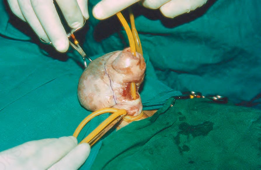
Fig. 12.282 Chronic edema simulating a keloid: note the dorsal proximal swelling and ventral urethral fistula. Courtesy of Dr Rameshwar Bang, Safat, Kuwait. Reproduced from Bang R.L. Penile edema induced by continuous condom catheter use and mimicking keloid scar. Scand J Urol Nephrol 1994;28:333–5. From Bunker C. Male Genital Skin Disease. Saunders Ltd./Elsevier 2004.
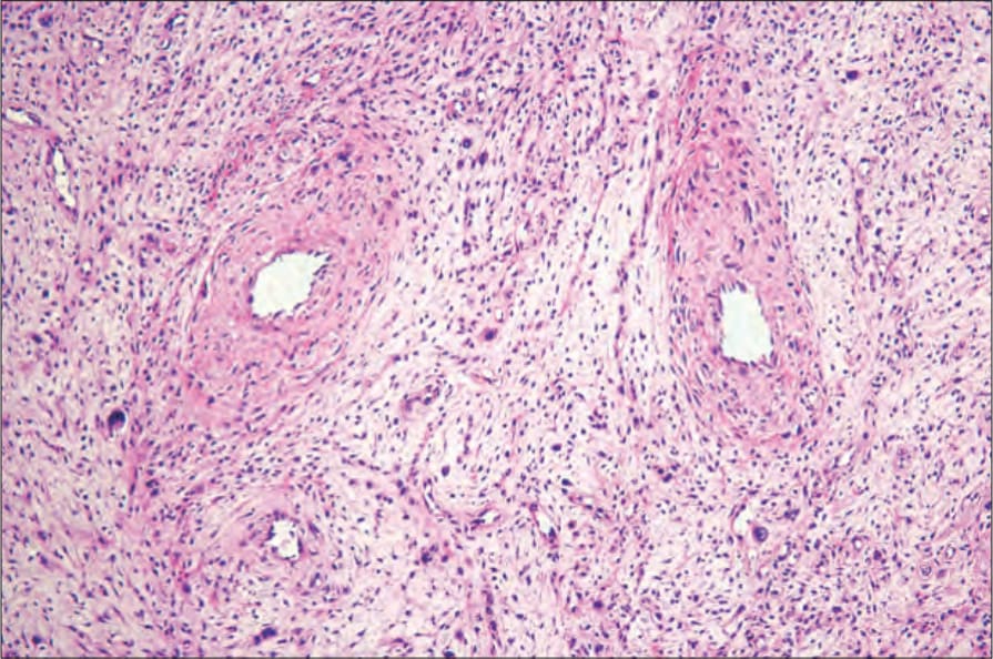
Fig. 12.283 Fibroepithelial stromal polyp: there are thick-walled vessels associated with a variably cellular loose connective tissue stroma. By courtesy of M. Nucci, MD, Brigham and Women’s Hospital and Harvard Medical School, Boston, USA.
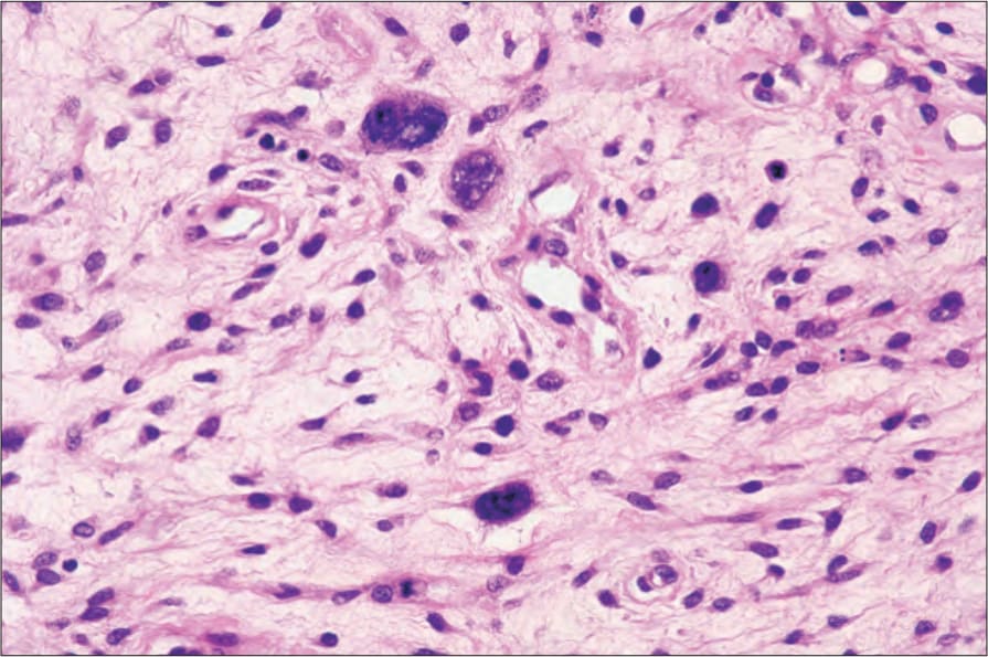
Fig. 12.284 Fibroepithelial stromal polyp: in this field, there is striking nuclear atypia. By courtesy of M. Nucci, MD, Brigham and Women’s Hospital and Harvard Medical School, Boston, USA.